The PLVAP Antibody (CAB15906) is a high-quality antibody developed for reliable detection and analysis of target proteins. This antibody, produced in rabbits, has high reactivity with human samples and is validated for use in Western blot applications. By binding to the PLVAP protein, researchers can investigate its role in various physiological and pathological conditions, particularly those related to vascular function and disease.PLVAP, also known as plasmalemmal vesicle-associated protein, is a key player in controlling the passage of molecules and cells across the endothelial barrier, making it a target of interest in studies on vascular biology, inflammation, and the development of therapies for conditions such as edema, tumor angiogenesis, and cardiovascular disease.
This antibody is validated for use in WB, ELISA applications and has demonstrated reactivity against Mouse, Rat samples.
Product Name:
PLVAP Antibody
SKU:
CAB15906
Size:
20μL, 100μL
Reactivity:
Mouse, Rat
Conjugate:
Unconjugated
Immunogen:
Recombinant protein (or fragment).This information is considered to be commercially sensitive.
Recommended starting concentration is 1 μg/mL. Please optimize the concentration based on your specific assay requirements.
Synonyms:
PV1, FELS, PV-1, gp68, DIAR10, PLVAP
Positive Sample:
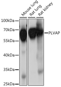
Mouse lung, Rat lung, Rat kidney
Cellular Localization:
Cell Membrane, Cytoplasm, Membrane, Single-Pass Type Ii Membrane Protein, Caveola, Perinuclear Region.
Calculated MW:
51kDa
Observed MW:
55kDa
Predicted to enable identical protein binding activity. Involved in MAPK cascade; positive regulation of cellular extravasation; and tumor necrosis factor-mediated signaling pathway. Located in cell surface. Colocalizes with caveola. Implicated in congenital diarrhea.
Purification Method
Affinity purification
Gene ID
83483
RRID
AB_2763337
Buffer Information
Store at -20℃. Avoid freeze / thaw cycles. Buffer: PBS with 0.01% thimerosal,50% glycerol,pH7.3.
Western blot analysis of various lysates using PLVAP Rabbit pAb (CAB15906) at 1:1000 dilution. Secondary antibody: HRP-conjugated Goat anti-Rabbit IgG (H+L) (CABS014) at 1:10000 dilution. Lysates/proteins: 25μg per lane. Blocking buffer: 3% nonfat dry milk in TBST. Detection: ECL Basic Kit (AbGn00020). Exposure time: 10s.