The PMAIP1 Antibody (CAB9801) is a high-quality antibody developed for reliable detection and analysis of target proteins. This antibody, generated in rabbits, is highly specific to human samples and is validated for use in Western blot applications. By binding to the PMAIP1 protein, this antibody allows for the detection and analysis of PMAIP1 expression in various cell types, making it ideal for studies in oncology and cell biology.PMAIP1, a pro-apoptotic protein, plays a crucial role in regulating programmed cell death by inducing mitochondrial membrane permeabilization. Its involvement in the intrinsic apoptosis pathway highlights its significance in cancer biology and potential as a therapeutic target.
This antibody is validated for use in WB, IF/ICC, ELISA, IF-P applications and has demonstrated reactivity against Human, Mouse, Rat samples.
Product Name:
PMAIP1 Antibody
SKU:
CAB9801
Size:
20μL, 100μL
Reactivity:
Human, Mouse, Rat
Conjugate:
Unconjugated
Immunogen:
Synthetic peptide. This information is considered to be commercially sensitive.
Recommended starting concentration is 1 μg/mL. Please optimize the concentration based on your specific assay requirements.
Synonyms:
APR, NOXA, PMAIP1
Positive Sample:
293F
Cellular Localization:
Mitochondrion.
Calculated MW:
6kDa
Observed MW:
15kDa
This gene belongs to a pro-apoptotic subfamily within the BCL-2 protein family, referred to as the BCL-2 homology domain 3 (BH3)-only subfamily, which determine whether a cell commits to apoptosis. In response to death-inducing stimuli, BH3-only members inhibit the anti-apoptotic BCL-2 family members, which under steady-state conditions keep the multi-BH domain proteins BAX and BAK, in an inactive state.
Purification Method
Affinity purification
Gene ID
5366
RRID
AB_2771718
Buffer Information
Store at -20℃. Avoid freeze / thaw cycles. Buffer: PBS containing 50% glycerol, preserved with proclin300 or sodium azide, pH 7.3.
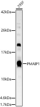
Western blot analysis of various lysates, using PMAIP1 Rabbit pAb (CAB9801) at 1:5000 dilution. Secondary antibody: HRP-conjugated Goat anti-Rabbit IgG (H+L) (CABS014) at 1:10000 dilution. Lysates/proteins: 25μg per lane. Blocking buffer: 3% nonfat dry milk in TBST. Detection: ECL Basic Kit (AbGn00020). Exposure time: 180s.
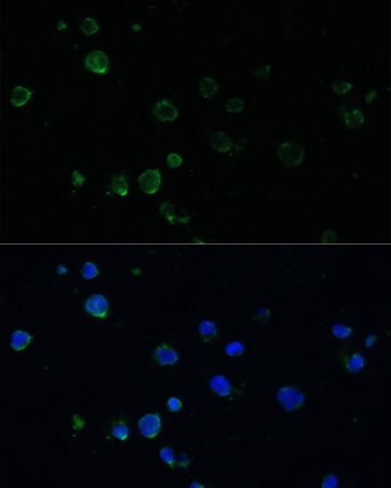
Immunofluorescence analysis of Jurkat cells using PMAIP1 Rabbit pAb (CAB9801) at dilution of 1:100. Secondary antibody: Cy3-conjugated Goat anti-Rabbit IgG (H+L) (CABS007) at 1:500 dilution. Blue: DAPI for nuclear staining.
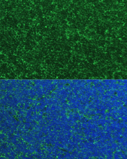
Immunofluorescence analysis of paraffin-embedded mouse thymus using PMAIP1 Rabbit pAb (CAB9801) at dilution of 1:100. Secondary antibody: Cy3-conjugated Goat anti-Rabbit IgG (H+L) (CABS007) at 1:500 dilution. Blue: DAPI for nuclear staining.