The CYPOR Monoclonal Antibody (CAB5032) is a high-quality antibody developed for reliable detection and analysis of target proteins. This antibody, generated in rabbits, exhibits high specificity for human samples and has been validated for use in Western blot applications. By binding to the CYPOR protein, this antibody enables accurate detection and analysis in a variety of cell types, making it invaluable for investigations in pharmacology and toxicology.CYPOR plays a crucial role in the activation of various drugs and xenobiotics by facilitating their metabolism in the liver.
This antibody is validated for use in WB, IHC-P, IF/ICC, ELISA applications and has demonstrated reactivity against Human, Mouse, Rat samples.
Product Name:
CYPOR Monoclonal Antibody
SKU:
CAB5032
Size:
20μL, 100μL
Reactivity:
Human, Mouse, Rat
Clone Number:
ARC1981
Conjugate:
Unconjugated
Immunogen:
Recombinant protein (or fragment).This information is considered to be commercially sensitive.
This gene encodes an endoplasmic reticulum membrane oxidoreductase that is essential for multiple metabolic processes, including reactions catalyzed by cytochrome P450 proteins for metabolism of steroid hormones, drugs and xenobiotics. The encoded protein has a flavin adenine dinucleotide (FAD)-binding domain and a flavodoxin-like domain which bind two cofactors, FAD and FMN, that allow it to donate electrons directly from NADPH to all microsomal P450 enzymes. Mutations in this gene cause a complex set of disorders, including apparent combined P450C17 and P450C21 deficiency, amenorrhea and disordered steroidogenesis, congenital adrenal hyperplasia and Antley-Bixler syndrome, that resemble those caused by defects in steroid metabolizing enzymes such as aromatase, 21-hydroxylase, and 17 alpha-hydroxylase.
Purification Method
Affinity purification
Gene ID
5447
RRID
AB_2863422
Buffer Information
Store at -20℃. Avoid freeze / thaw cycles. Buffer: PBS containing 50% glycerol and 0.05% BSA, preserved with proclin300 or sodium azide, pH 7.3.
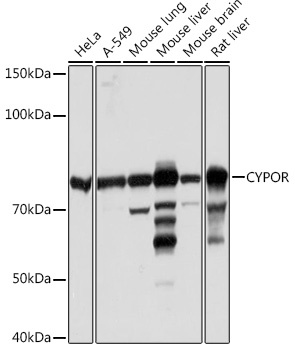
Western blot analysis of various lysates using CYPOR Rabbit mAb (CAB5032) at 1:1000 dilution. Secondary antibody: HRP-conjugated Goat anti-Rabbit IgG (H+L) (CABS014) at 1:10000 dilution. Lysates/proteins: 25μg per lane. Blocking buffer: 3% nonfat dry milk in TBST. Detection: ECL Enhanced Kit (AbGn00021). Exposure time: 10s.
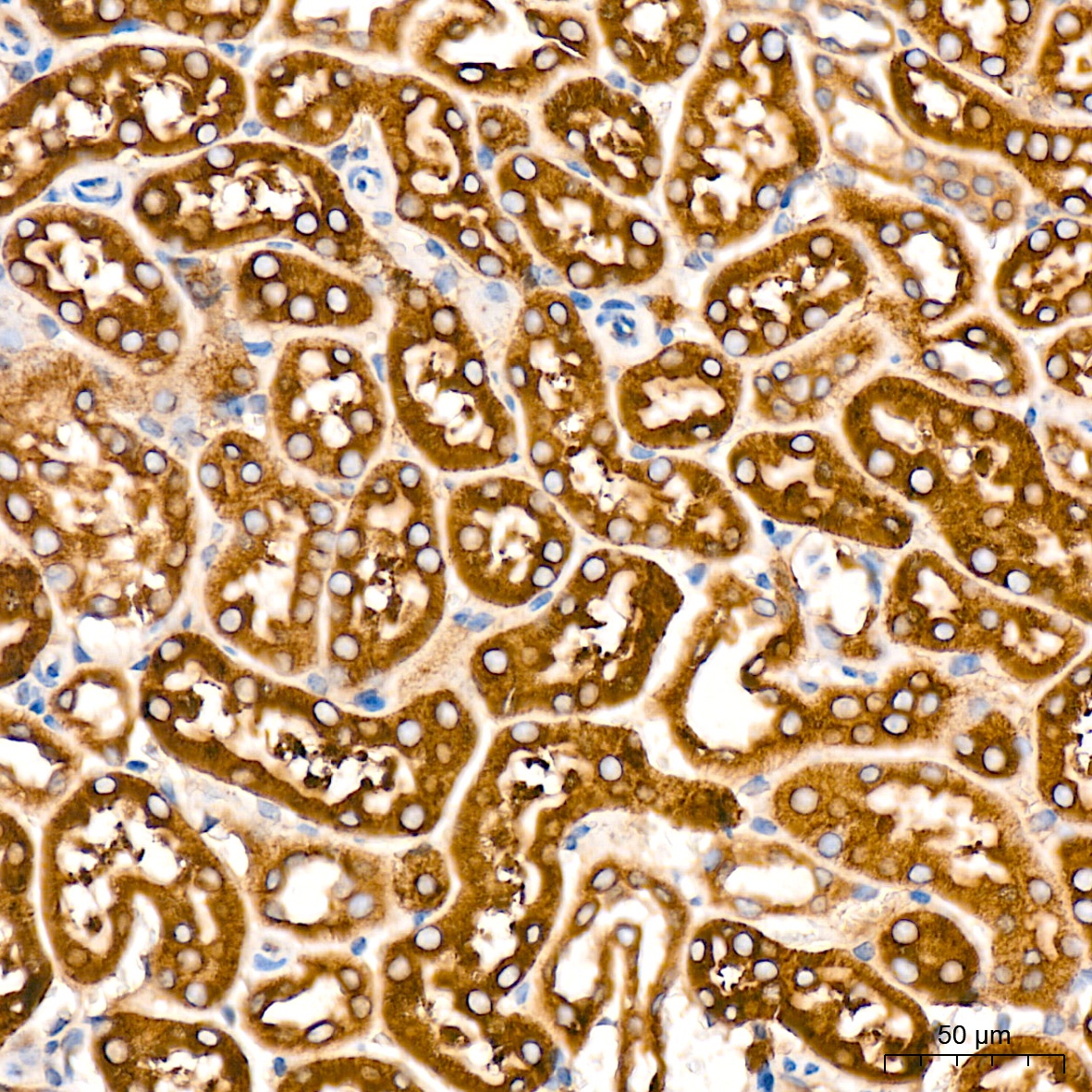
Immunohistochemistry analysis of paraffin-embedded Mouse kidney tissue using CYPOR Rabbit mAb (CAB5032) at a dilution of 1:200 (40x lens). High pressure antigen retrieval was performed with 0.01 M citrate buffer (pH 6.0) prior to IHC staining.
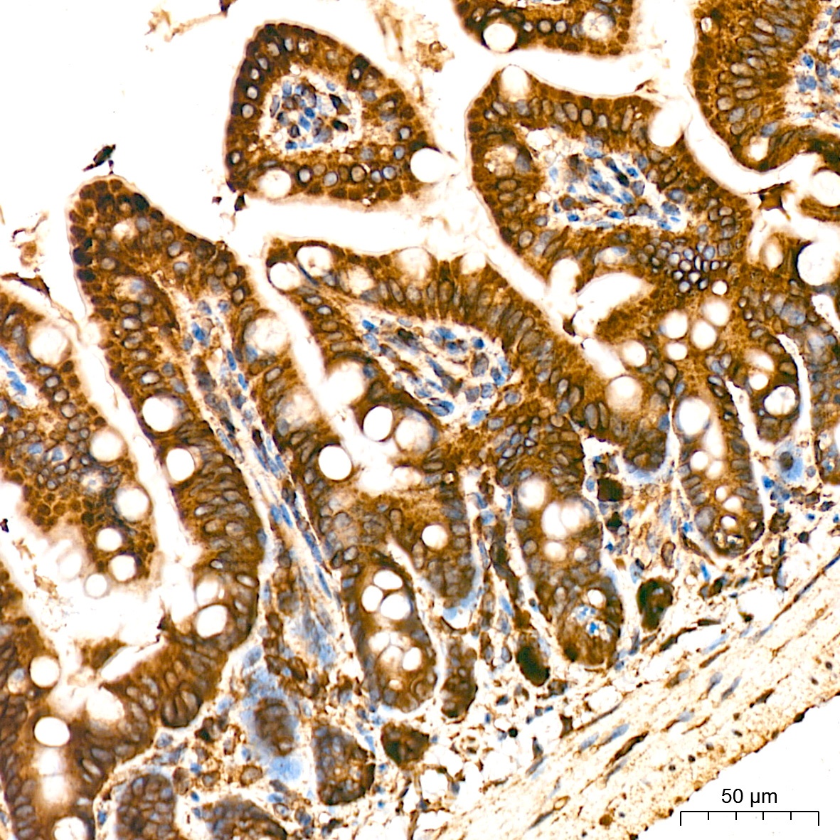
Immunohistochemistry analysis of paraffin-embedded Mouse intestin tissue using CYPOR Rabbit mAb (CAB5032) at a dilution of 1:200 (40x lens). High pressure antigen retrieval was performed with 0.01 M citrate buffer (pH 6.0) prior to IHC staining.
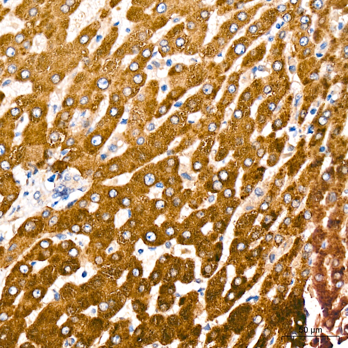
Immunohistochemistry analysis of paraffin-embedded Human liver tissue using CYPOR Rabbit mAb (CAB5032) at a dilution of 1:200 (40x lens). High pressure antigen retrieval was performed with 0.01 M citrate buffer (pH 6.0) prior to IHC staining.
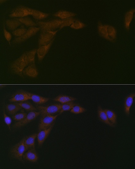
Immunofluorescence analysis of U-2 OS cells using CYPOR Rabbit mAb (CAB5032) at dilution of 1:100 (40x lens). Secondary antibody: Cy3-conjugated Goat anti-Rabbit IgG (H+L) (CABS007) at 1:500 dilution. Blue: DAPI for nuclear staining.


")


