The PPM1A Antibody (CAB6699) is a high-quality antibody developed for reliable detection and analysis of target proteins. This antibody, produced in rabbits, exhibits high reactivity with human samples and is optimized for use in Western blot applications. By binding to the PPM1A protein, this antibody enables accurate detection and analysis in a variety of cell types, making it ideal for investigations in cell biology, cancer research, and drug development.PPM1A, also known as protein phosphatase Mg2+/Mn2+ dependent 1A, is involved in dephosphorylation events that control key signaling pathways crucial for cellular function and survival.
This antibody is validated for use in WB, IF/ICC, ELISA applications and has demonstrated reactivity against Human, Mouse, Rat samples.
Product Name:
PPM1A Antibody
SKU:
CAB6699
Size:
20μL, 100μL
Reactivity:
Human, Mouse, Rat
Conjugate:
Unconjugated
Immunogen:
Recombinant protein (or fragment).This information is considered to be commercially sensitive.
The protein encoded by this gene is a member of the PP2C family of Ser/Thr protein phosphatases. PP2C family members are known to be negative regulators of cell stress response pathways. This phosphatase dephosphorylates, and negatively regulates the activities of, MAP kinases and MAP kinase kinases. It has been shown to inhibit the activation of p38 and JNK kinase cascades induced by environmental stresses. This phosphatase can also dephosphorylate cyclin-dependent kinases, and thus may be involved in cell cycle control. Overexpression of this phosphatase is reported to activate the expression of the tumor suppressor gene TP53/p53, which leads to G2/M cell cycle arrest and apoptosis. Three alternatively spliced transcript variants encoding distinct isoforms have been described.
Purification Method
Affinity purification
Gene ID
5494
RRID
AB_2767283
Buffer Information
Store at -20℃. Avoid freeze / thaw cycles. Buffer: PBS containing 50% glycerol, preserved with proclin300 or sodium azide, pH 7.3.
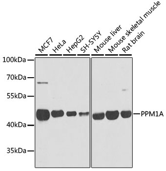
Western blot analysis of various lysates using PPM1A Rabbit pAb (CAB6699) at 1:1000 dilution. Secondary antibody: HRP-conjugated Goat anti-Rabbit IgG (H+L) (CABS014) at 1:10000 dilution. Lysates/proteins: 25μg per lane. Blocking buffer: 3% nonfat dry milk in TBST. Detection: ECL Basic Kit (AbGn00020). Exposure time: 1s.
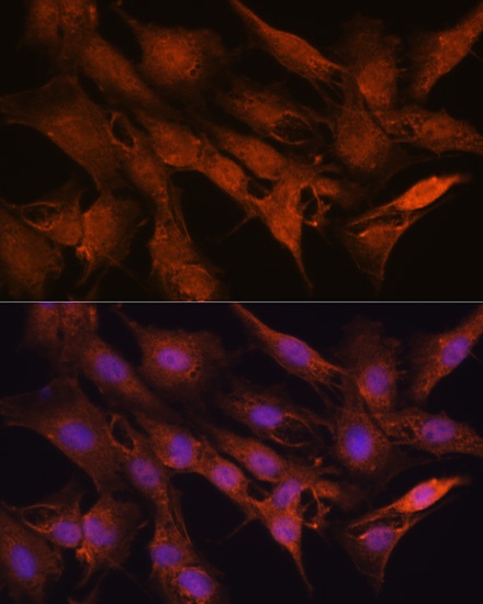
Immunofluorescence analysis of C6 cells using PPM1A Rabbit pAb (CAB6699) at dilution of 1:100 (40x lens). Secondary antibody: Cy3-conjugated Goat anti-Rabbit IgG (H+L) (CABS007) at 1:500 dilution. Blue: DAPI for nuclear staining.
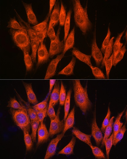
Immunofluorescence analysis of NIH-3T3 cells using PPM1A Rabbit pAb (CAB6699) at dilution of 1:100 (40x lens). Secondary antibody: Cy3-conjugated Goat anti-Rabbit IgG (H+L) (CABS007) at 1:500 dilution. Blue: DAPI for nuclear staining.