The PPP1R15A Antibody (CAB16260) is a high-quality antibody developed for reliable detection and analysis of target proteins. This antibody, raised in rabbits, exhibits high reactivity with human samples and has been validated for use in Western blot applications. By specifically binding to the PPP1R15A protein, this antibody allows for accurate detection and analysis in a variety of cell types, making it ideal for studies in cell biology and molecular biology research.PPP1R15A, also known as GADD34, is a key player in the unfolded protein response pathway, which is activated in response to cellular stress such as endoplasmic reticulum (ER) stress.
This antibody is validated for use in WB, IF/ICC, ELISA applications and has demonstrated reactivity against Human, Mouse, Rat samples.
Product Name:
PPP1R15A Antibody
SKU:
CAB16260
Size:
20μL, 100μL
Reactivity:
Human, Mouse, Rat
Immunogen:
Recombinant protein (or fragment).This information is considered to be commercially sensitive.
Cytoplasm, Cytosol, Endoplasmic Reticulum, Golgi Apparatus, Mitochondrial Outer Membrane, Mitochondrion, Protein Phosphatase Type 1 Complex.
Calculated MW:
73kDa
Observed MW:
110kDa
This gene is a member of a group of genes whose transcript levels are increased following stressful growth arrest conditions and treatment with DNA-damaging agents. The induction of this gene by ionizing radiation occurs in certain cell lines regardless of p53 status, and its protein response is correlated with apoptosis following ionizing radiation.
Purification Method
Affinity purification
Gene ID
23645
RRID
AB_2771772
Buffer Information
Store at -20℃. Avoid freeze / thaw cycles. Buffer: PBS with 0.01% thimerosal,50% glycerol,pH7.3.
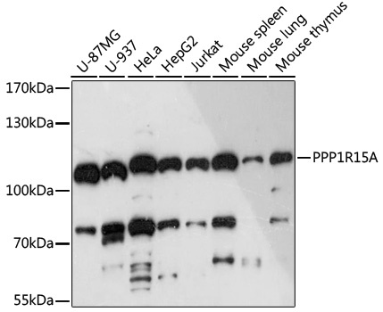
Western blot analysis of various lysates using PPP1R15A Rabbit pAb (CAB16260) at 1:3000 dilution. Secondary antibody: HRP-conjugated Goat anti-Rabbit IgG (H+L) (CABS014) at 1:10000 dilution. Lysates/proteins: 25μg per lane. Blocking buffer: 3% nonfat dry milk in TBST. Detection: ECL Basic Kit (AbGn00020). Exposure time: 90s.
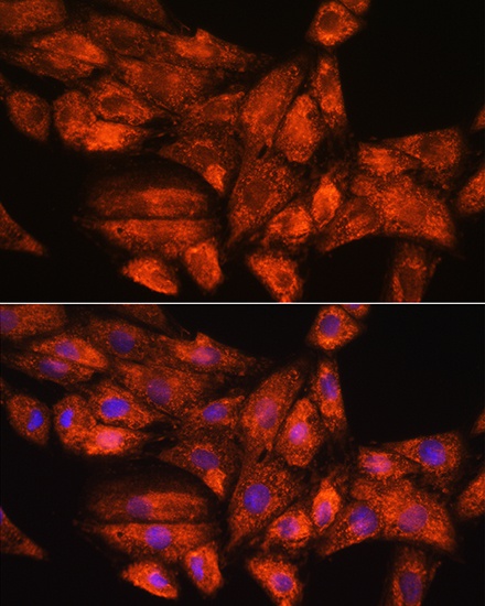
Immunofluorescence analysis of H9C2 cells using PPP1R15A Rabbit pAb (CAB16260) at dilution of 1:100. Secondary antibody: Cy3-conjugated Goat anti-Rabbit IgG (H+L) (CABS007) at 1:500 dilution. Blue: DAPI for nuclear staining.