The PRC1 Antibody (CAB7029) is a high-quality antibody developed for reliable detection and analysis of target proteins. This antibody, generated in rabbits, has been optimized for use in Western blot applications and demonstrates high specificity and sensitivity when detecting PRC1 in human samples.PRC1 plays a vital role in ensuring accurate chromosome segregation during cell division, making it a key target for research in cancer biology and cell cycle regulation. Dysregulation of PRC1 expression or function has been associated with various types of cancer, making it an important focus for understanding tumor development and progression.
This antibody is validated for use in WB, IHC-P, IF/ICC, ELISA applications and has demonstrated reactivity against Human, Mouse, Rat samples.
Product Name:
PRC1 Antibody
SKU:
CAB7029
Size:
20μL, 100μL
Reactivity:
Human, Mouse, Rat
Conjugate:
Unconjugated
Immunogen:
Recombinant protein (or fragment).This information is considered to be commercially sensitive.
Recommended starting concentration is 1 μg/mL. Please optimize the concentration based on your specific assay requirements.
Synonyms:
ASE1, PRC1
Positive Sample:
MCF7, Jurkat, 293T, HepG2
Cellular Localization:
Cytoplasm, Midbody, Nucleus, Cytoskeleton, Spindle Pole.
Calculated MW:
72kDa
Observed MW:
72kDa
This gene encodes a protein that is involved in cytokinesis. The protein is present at high levels during the S and G2/M phases of mitosis but its levels drop dramatically when the cell exits mitosis and enters the G1 phase. It is located in the nucleus during interphase, becomes associated with mitotic spindles in a highly dynamic manner during mitosis, and localizes to the cell mid-body during cytokinesis. This protein has been shown to be a substrate of several cyclin-dependent kinases (CDKs). It is necessary for polarizing parallel microtubules and concentrating the factors responsible for contractile ring assembly. Alternative splicing results in multiple transcript variants.
Purification Method
Affinity purification
Gene ID
9055
RRID
AB_2767584
Buffer Information
Store at -20℃. Avoid freeze / thaw cycles. Buffer: PBS containing 50% glycerol, preserved with proclin300 or sodium azide, pH 7.3.
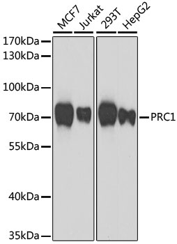
Western blot analysis of various lysates using PRC1 Rabbit pAb (CAB7029) at 1:1000 dilution. Secondary antibody: HRP-conjugated Goat anti-Rabbit IgG (H+L) (CABS014) at 1:10000 dilution. Lysates/proteins: 25μg per lane. Blocking buffer: 3% nonfat dry milk in TBST. Detection: ECL Basic Kit (AbGn00020). Exposure time: 90s.
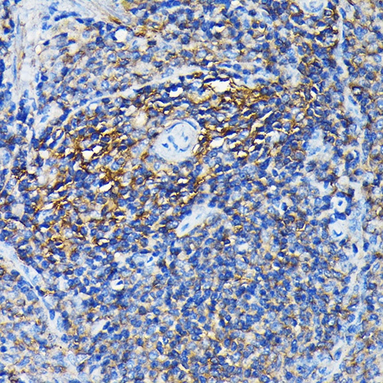
Immunohistochemistry analysis of paraffin-embedded Rat spleen using PRC1 Rabbit pAb (CAB7029) at dilution of 1:100 (40x lens). High pressure antigen retrieval performed with 0.01M Citrate buffer (pH 6.0) prior to IHC staining.
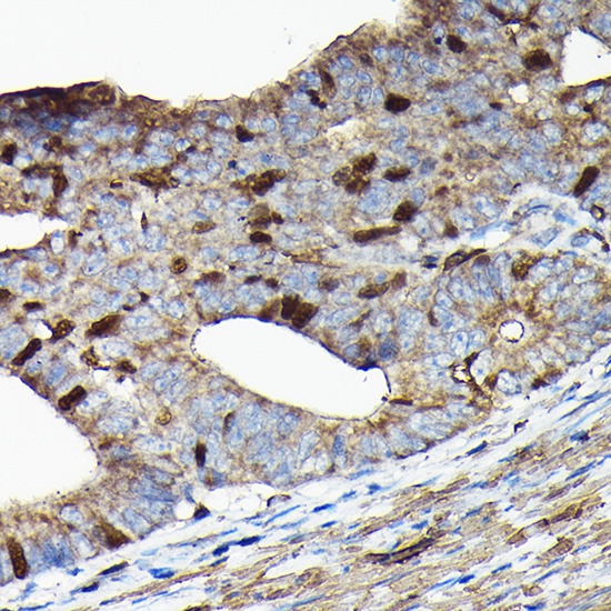
Immunohistochemistry analysis of paraffin-embedded Human colon carcinoma using PRC1 Rabbit pAb (CAB7029) at dilution of 1:100 (40x lens). High pressure antigen retrieval performed with 0.01M Citrate buffer (pH 6.0) prior to IHC staining.
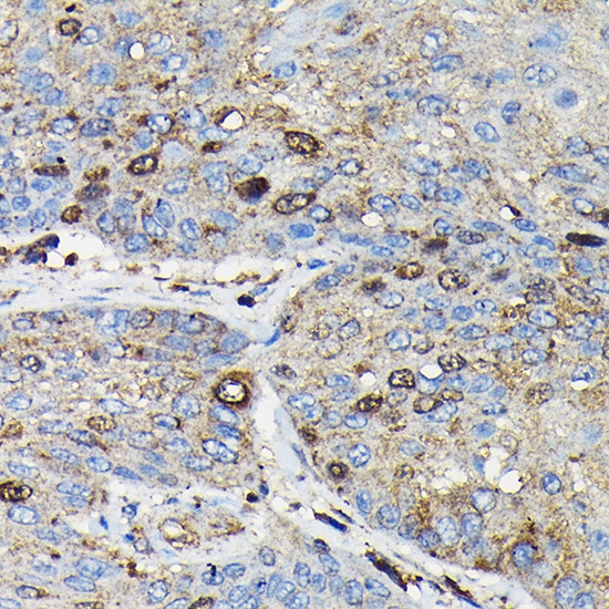
Immunohistochemistry analysis of paraffin-embedded Human esophageal cancer using PRC1 Rabbit pAb (CAB7029) at dilution of 1:100 (40x lens). High pressure antigen retrieval performed with 0.01M Citrate buffer (pH 6.0) prior to IHC staining.
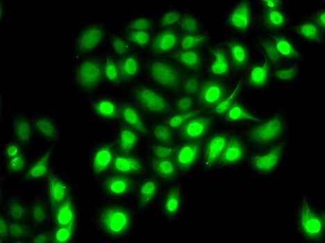
Immunofluorescence analysis of MCF7 cells using PRC1 Rabbit pAb (CAB7029).Secondary antibody: Cy3-conjugated Goat anti-Rabbit IgG (H+L) (CABS007) at 1:500 dilution.