The NGFRAP1 Antibody (CAB7296) is a high-quality antibody developed for reliable detection and analysis of target proteins. This antibody, produced in rabbits, demonstrates high reactivity with human samples and is validated for use in Western blot applications. By specifically binding to the BEX3 protein, this antibody allows for accurate detection and analysis in various cell types, making it ideal for studies in neuroscience and neurodegenerative diseases.BEX3 is known for its role in promoting neuronal differentiation and survival, and its dysregulation has been implicated in various neurological disorders, including Alzheimer's disease and Parkinson's disease.
This antibody is validated for use in WB, IF/ICC, ELISA applications and has demonstrated reactivity against Human, Mouse samples.
Product Name:
NGFRAP1 Antibody
SKU:
CAB7296
Size:
20μL, 100μL
Reactivity:
Human, Mouse
Conjugate:
Unconjugated
Immunogen:
Recombinant protein (or fragment).This information is considered to be commercially sensitive.
Recommended starting concentration is 1 μg/mL. Please optimize the concentration based on your specific assay requirements.
Synonyms:
Bex, NADE, HGR74, NGFRAP1, DXS6984E
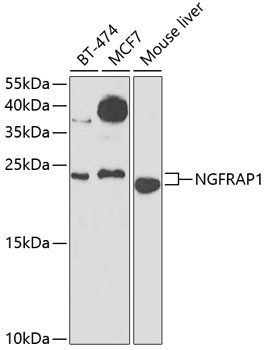
Positive Sample:
BT-474, MCF7, Mouse liver
Cellular Localization:
Cytoplasm, Nucleus.
Calculated MW:
13kDa
Observed MW:
24kDa
Enables identical protein binding activity. Predicted to be involved in signal transduction. Predicted to act upstream of or within extrinsic apoptotic signaling pathway via death domain receptors. Located in cytosol.
Purification Method
Affinity purification
Gene ID
27018
RRID
AB_2767837
Buffer Information
Store at -20℃. Avoid freeze / thaw cycles. Buffer: PBS containing 50% glycerol, preserved with proclin300 or sodium azide, pH 7.3.
Western blot analysis of various lysates using NGFRAP1 Rabbit pAb (CAB7296) at 1:1000 dilution. Secondary antibody: HRP-conjugated Goat anti-Rabbit IgG (H+L) (CABS014) at 1:10000 dilution. Lysates/proteins: 25μg per lane. Blocking buffer: 3% nonfat dry milk in TBST. Detection: ECL Enhanced Kit (AbGn00021). Exposure time: 90s.
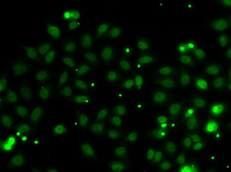
Immunofluorescence analysis of MCF-7 cells using NGFRAP1 Rabbit pAb (CAB7296).Secondary antibody: Cy3-conjugated Goat anti-Rabbit IgG (H+L) (CABS007) at 1:500 dilution.