The PP1 beta Monoclonal Antibody (CAB4364) is a high-quality antibody developed for reliable detection and analysis of target proteins. This antibody, raised in rabbits, is highly specific and reactive with human samples, providing reliable results in Western blot applications.Protein Phosphatase 1 Beta is involved in the dephosphorylation of various proteins, impacting processes such as cell cycle progression, gene expression, and metabolism. Dysregulation of this enzyme has been linked to diseases like cancer, diabetes, and neurodegenerative disorders, making it a valuable target for therapeutic interventions.
This antibody is validated for use in WB, IHC-P, IF/ICC, ELISA applications and has demonstrated reactivity against Human, Mouse, Rat samples.
Product Name:
PP1 beta Monoclonal Antibody
SKU:
CAB4364
Size:
20μL, 100μL
Reactivity:
Human, Mouse, Rat
Clone Number:
ARC0981
Conjugate:
Unconjugated
Immunogen:
Synthetic peptide. This information is considered to be commercially sensitive.
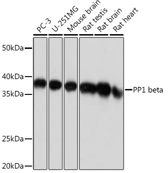
PC-3, U-251MG, Mouse brain, Rat testis, Rat brain, Rat heart
Cellular Localization:
Cytoplasm, Nucleus, Nucleolus, Nucleoplasm.
Calculated MW:
37kDa
Observed MW:
37kDa
The protein encoded by this gene is one of the three catalytic subunits of protein phosphatase 1 (PP1). PP1 is a serine/threonine specific protein phosphatase known to be involved in the regulation of a variety of cellular processes, such as cell division, glycogen metabolism, muscle contractility, protein synthesis, and HIV-1 viral transcription. Mouse studies suggest that PP1 functions as a suppressor of learning and memory. Two alternatively spliced transcript variants encoding distinct isoforms have been observed.
Purification Method
Affinity purification
Gene ID
5500
RRID
AB_2863249
Buffer Information
Store at -20℃. Avoid freeze / thaw cycles. Buffer: PBS containing 50% glycerol and 0.05% BSA, preserved with proclin300 or sodium azide, pH 7.3.
Western blot analysis of various lysates using PP1 beta Rabbit mAb (CAB4364) at 1:1000 dilution. Secondary antibody: HRP-conjugated Goat anti-Rabbit IgG (H+L) (CABS014) at 1:10000 dilution. Lysates/proteins: 25μg per lane. Blocking buffer: 3% nonfat dry milk in TBST. Detection: ECL Basic Kit (AbGn00020). Exposure time: 1s.
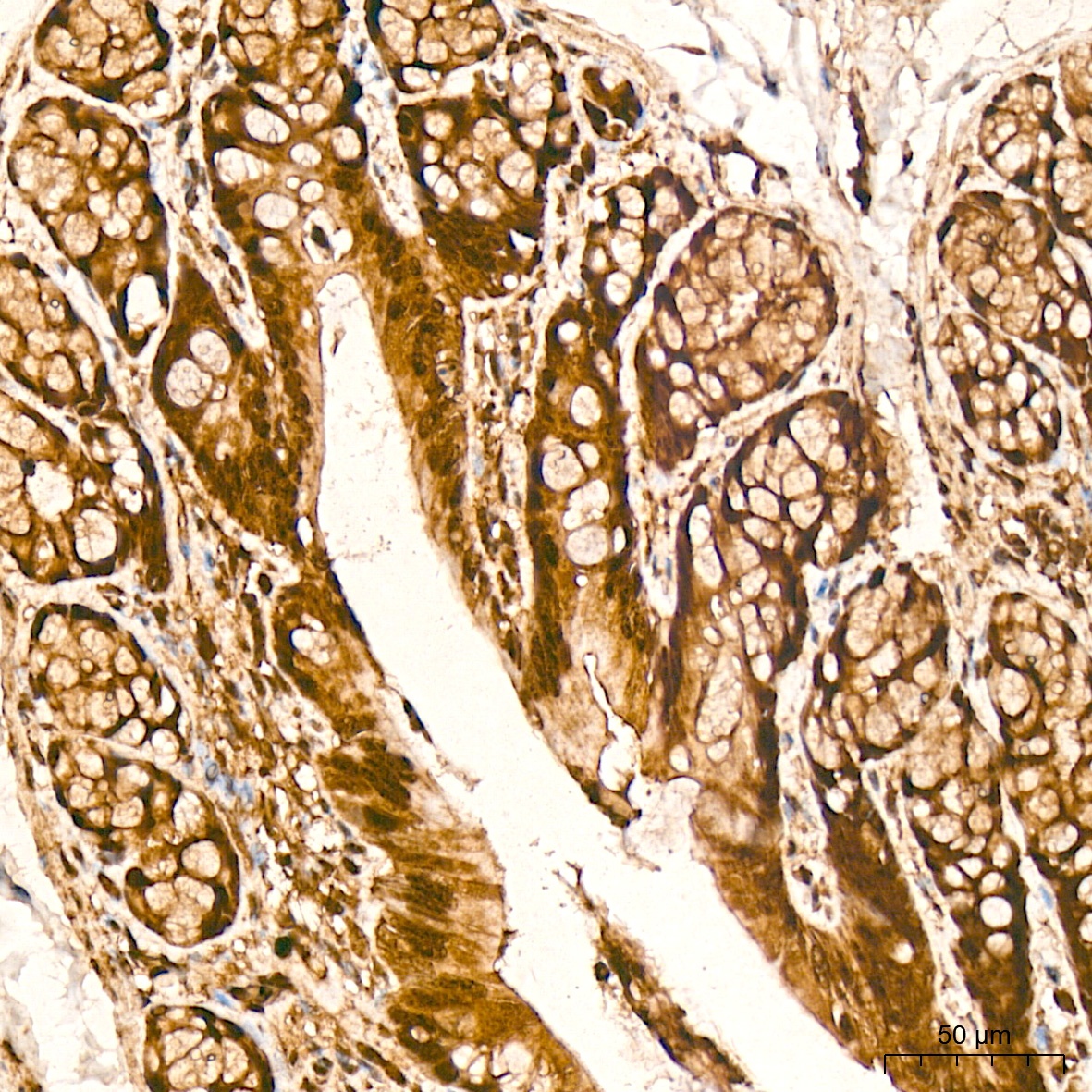
Immunohistochemistry analysis of paraffin-embedded Rat colon tissue using PP1 beta Rabbit mAb (CAB4364) at a dilution of 1:1000 (40x lens). High pressure antigen retrieval was performed with 0.01 M citrate buffer (pH 6.0) prior to IHC staining.
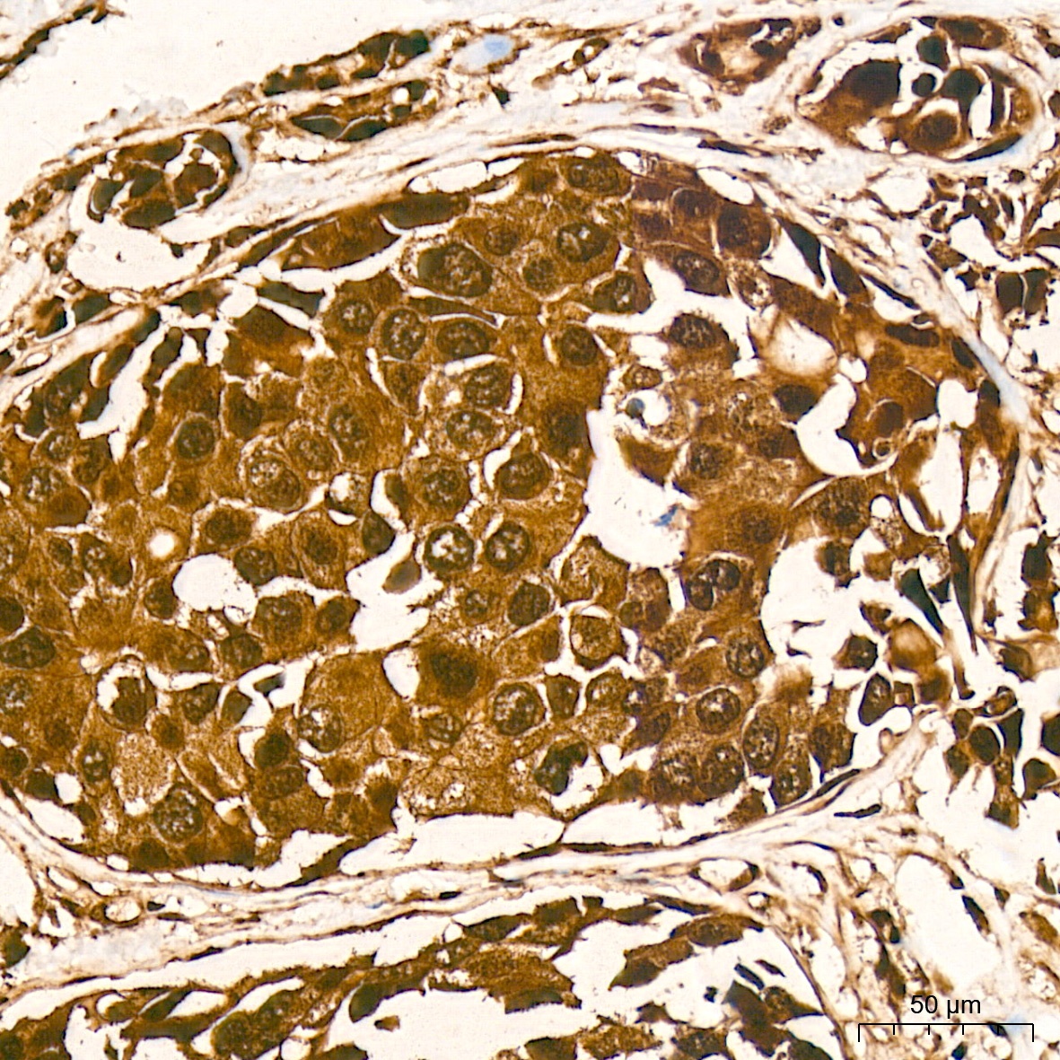
Immunohistochemistry analysis of paraffin-embedded Human breast cancer tissue using PP1 beta Rabbit mAb (CAB4364) at a dilution of 1:1000 (40x lens). High pressure antigen retrieval was performed with 0.01 M citrate buffer (pH 6.0) prior to IHC staining.
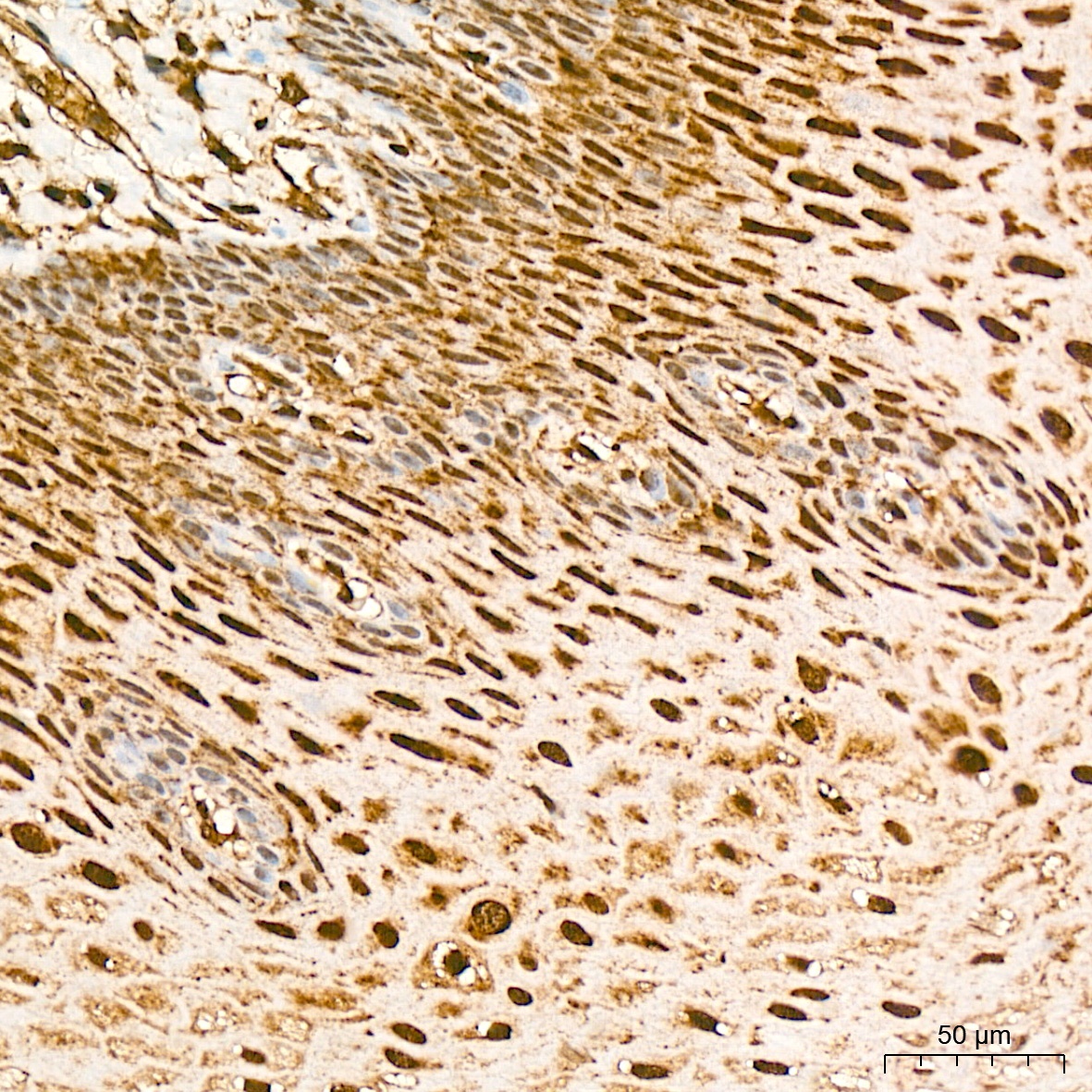
Immunohistochemistry analysis of paraffin-embedded Human esophagus tissue using PP1 beta Rabbit mAb (CAB4364) at a dilution of 1:1000 (40x lens). High pressure antigen retrieval was performed with 0.01 M citrate buffer (pH 6.0) prior to IHC staining.
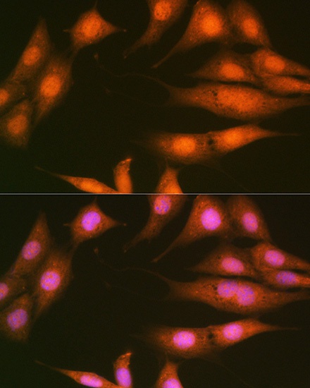
Immunofluorescence analysis of NIH-3T3 cells using PP1 beta Rabbit mAb (CAB4364) at dilution of 1:100 (40x lens). Secondary antibody: Cy3-conjugated Goat anti-Rabbit IgG (H+L) (CABS007) at 1:500 dilution. Blue: DAPI for nuclear staining.
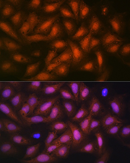
Immunofluorescence analysis of U-2 OS cells using PP1 beta Rabbit mAb (CAB4364) at dilution of 1:100 (40x lens). Secondary antibody: Cy3-conjugated Goat anti-Rabbit IgG (H+L) (CABS007) at 1:500 dilution. Blue: DAPI for nuclear staining.