The PSMA/FOLH1 Monoclonal Antibody (CAB22212) is a high-quality antibody developed for reliable detection and analysis of target proteins. This antibody, generated in mice, is highly specific and reactive with human samples, making it ideal for use in various research applications.PSMA/FOLH1 is a transmembrane protein that is highly expressed in prostate cancer cells, making it a promising target for cancer research and therapy development. The antibody binds specifically to the PSMA/FOLH1 protein, enabling accurate detection and analysis in a variety of cell types.
This antibody is validated for use in WB, IHC-P, ELISA applications and has demonstrated reactivity against Human, Mouse, Rat samples.
Product Name:
PSMA/FOLH1 Monoclonal Antibody
SKU:
CAB22212
Size:
20μL, 100μL
Reactivity:
Human, Mouse, Rat
Clone Number:
ARC56326
Conjugate:
Unconjugated
Immunogen:
Synthetic peptide. This information is considered to be commercially sensitive.
This gene encodes a type II transmembrane glycoprotein belonging to the M28 peptidase family. The protein acts as a glutamate carboxypeptidase on different alternative substrates, including the nutrient folate and the neuropeptide N-acetyl-l-aspartyl-l-glutamate and is expressed in a number of tissues such as prostate, central and peripheral nervous system and kidney. A mutation in this gene may be associated with impaired intestinal absorption of dietary folates, resulting in low blood folate levels and consequent hyperhomocysteinemia. Expression of this protein in the brain may be involved in a number of pathological conditions associated with glutamate excitotoxicity. In the prostate the protein is up-regulated in cancerous cells and is used as an effective diagnostic and prognostic indicator of prostate cancer. This gene likely arose from a duplication event of a nearby chromosomal region. Alternative splicing gives rise to multiple transcript variants encoding several different isoforms.
Purification Method
Affinity purification
Gene ID
2346
Buffer Information
Store at -20℃. Avoid freeze / thaw cycles. Buffer: PBS with 0.09% sodium azide,0.05% BSA,50% glycerol,pH7.3.
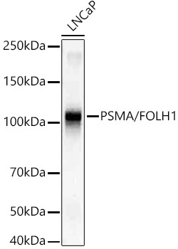
Western blot analysis of lysates from LNCaP cells, using PSMA/FOLH1 Rabbit mAb (CAB22212) at1:20000 dilution. Secondary antibody: HRP-conjugated Goat anti-Rabbit IgG (H+L) (CABS014) at 1:10000 dilution. Lysates/proteins: 25μg per lane. Blocking buffer: 3% nonfat dry milk in TBST. Detection: ECL Basic Kit (AbGn00020). Exposure time: 1s.
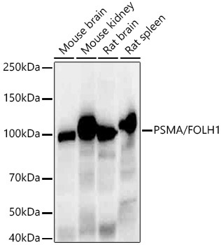
Western blot analysis of various lysates, using PSMA/FOLH1 Rabbit mAb (CAB22212) at1:20000 dilution. Secondary antibody: HRP-conjugated Goat anti-Rabbit IgG (H+L) (CABS014) at 1:10000 dilution. Lysates/proteins: 25μg per lane. Blocking buffer: 3% nonfat dry milk in TBST. Detection: ECL Enhanced Kit (AbGn00021). Exposure time: 30s.
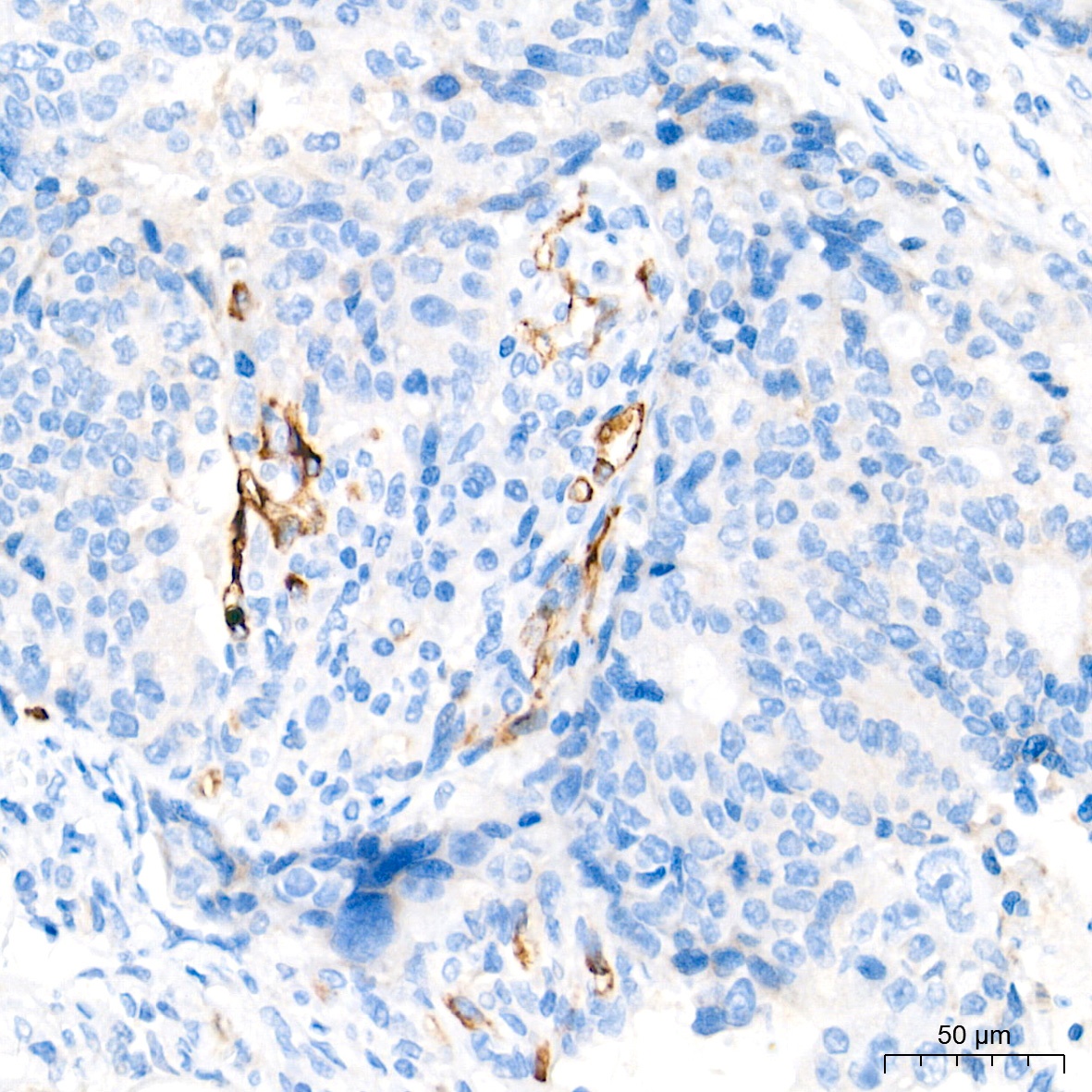
Immunohistochemistry analysis of paraffin-embedded Human colon carcinoma tissue using PSMA/FOLH1 Rabbit mAb (CAB22212) at a dilution of 1:15000 (40x lens). High pressure antigen retrieval performed with 0.01M Tris-EDTA Buffer (pH 9.0) prior to IHC staining.
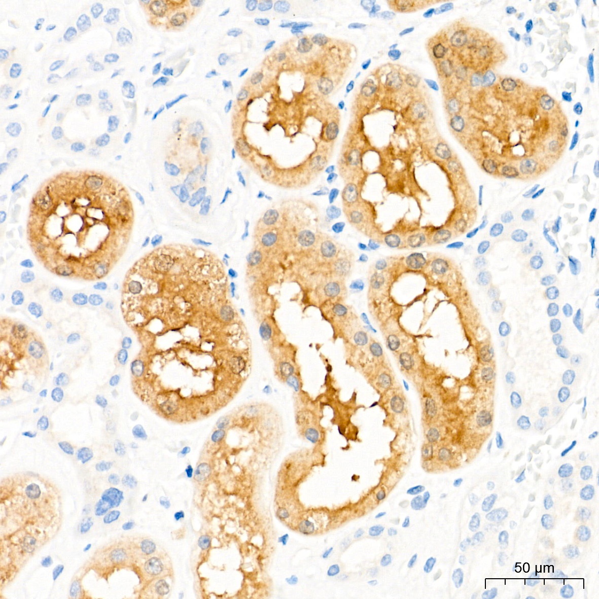
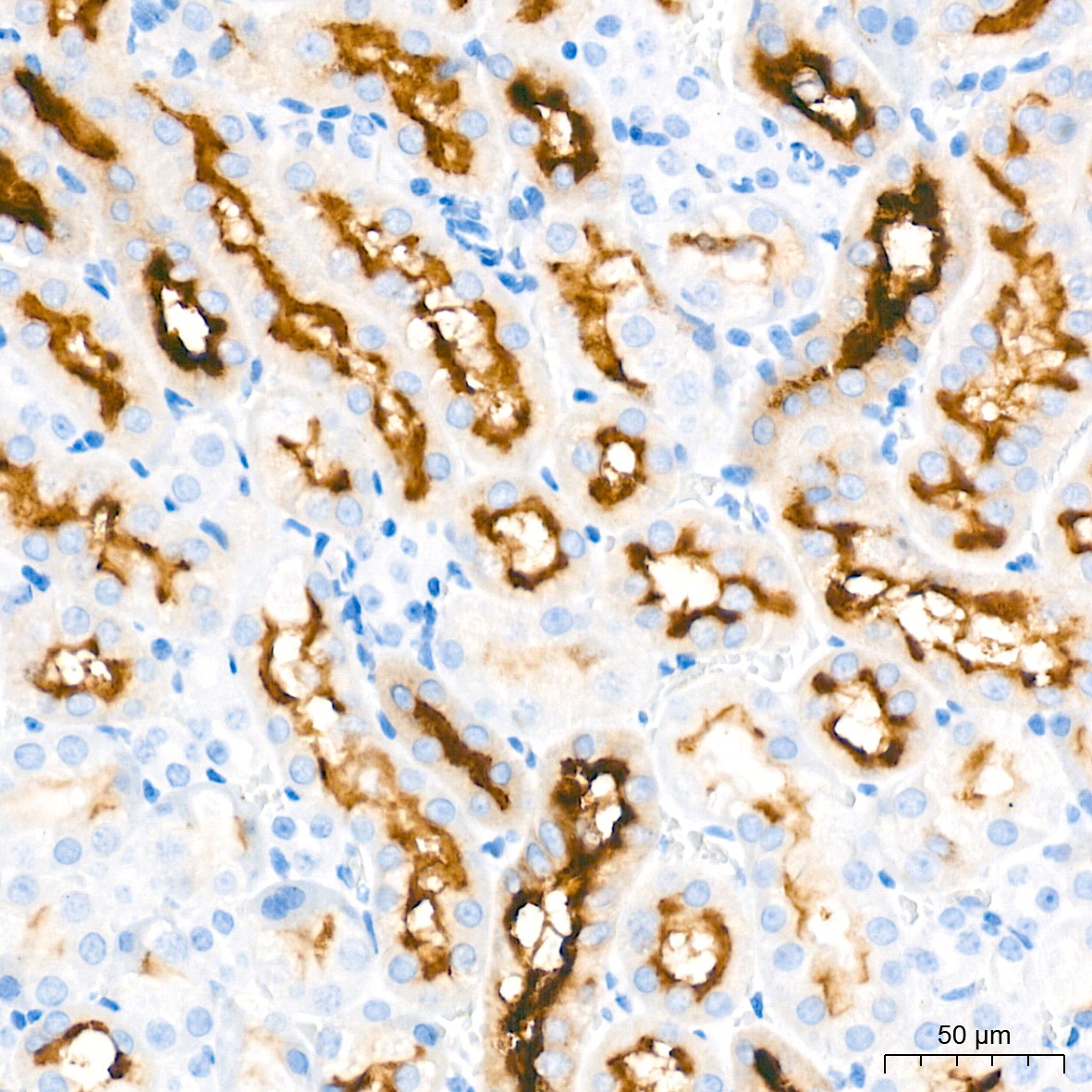
Immunohistochemistry analysis of paraffin-embedded Human kidney tissue using PSMA/FOLH1 Rabbit mAb (CAB22212) at a dilution of 1:15000 (40x lens). High pressure antigen retrieval performed with 0.01M Tris-EDTA Buffer (pH 9.0) prior to IHC staining.
Immunohistochemistry analysis of paraffin-embedded Mouse kidney tissue using PSMA/FOLH1 Rabbit mAb (CAB22212) at a dilution of 1:15000 (40x lens). High pressure antigen retrieval performed with 0.01M Tris-EDTA Buffer (pH 9.0) prior to IHC staining.
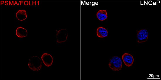
Confocal imaging of LNCaP cells using PSMA/FOLH1 Rabbit mAb (CAB22212, dilution 1:200) followed by a further incubation with Cy3-conjugated Goat anti-Rabbit IgG (H+L) (CABS007, dilution 1:500) (Red). DAPI was used for nuclear staining (Blue). Objective: 100x.
 at1:20000 dilution. Secondary antibody: HRP Goat Anti-Rabbit IgG (H+L) at 1:2000000 dilution. Lysates/proteins: 25μg per lane. Blocking buffer: 3% nonfat dry milk in TBST.")
 at1:20000 dilution. Secondary antibody: HRP Goat Anti-Rabbit IgG (H+L) at 1:2000000 dilution. Lysates/proteins: 25μg per lane. Blocking buffer: 3% nonfat dry milk in TBST.")

")

")
")