The PSMA6 Antibody (CAB13536) is a high-quality antibody developed for reliable detection and analysis of target proteins. The antibody is raised in rabbits and is highly reactive with human samples, making it suitable for a variety of research applications, including Western blot analysis. By binding specifically to the PSMA6 protein, this antibody enables the detection and analysis of PSMA6 in different cell types.PSMA6 is a key component of the proteasome complex, which plays a crucial role in degrading unwanted or damaged proteins within cells. Dysregulation of proteasome activity has been implicated in various diseases, including cancer, neurodegenerative disorders, and inflammatory conditions.
This antibody is validated for use in WB, IF/ICC, ELISA applications and has demonstrated reactivity against Human, Mouse, Rat samples.
Product Name:
PSMA6 Antibody
SKU:
CAB13536
Size:
20μL, 100μL
Reactivity:
Human, Mouse, Rat
Conjugate:
Unconjugated
Immunogen:
Recombinant protein (or fragment).This information is considered to be commercially sensitive.
The proteasome is a multicatalytic proteinase complex with a highly ordered ring-shaped 20S core structure. The core structure is composed of 4 rings of 28 non-identical subunits; 2 rings are composed of 7 alpha subunits and 2 rings are composed of 7 beta subunits. Proteasomes are distributed throughout eukaryotic cells at a high concentration and cleave peptides in an ATP/ubiquitin-dependent process in a non-lysosomal pathway. An essential function of a modified proteasome, the immunoproteasome, is the processing of class I MHC peptides. This gene encodes a member of the peptidase T1A family, that is a 20S core alpha subunit. Multiple transcript variants encoding several different isoforms have been found for this gene. A pseudogene has been identified on the Y chromosome.
Purification Method
Affinity purification
Gene ID
5687
RRID
AB_2760396
Buffer Information
Store at -20℃. Avoid freeze / thaw cycles. Buffer: PBS containing 50% glycerol, preserved with proclin300 or sodium azide, pH 7.3.
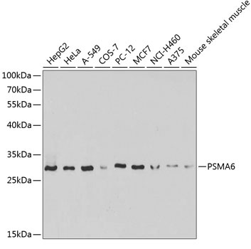
Western blot analysis of various lysates using PSMA6 Rabbit pAb (CAB13536) at 1:1000 dilution. Secondary antibody: HRP-conjugated Goat anti-Rabbit IgG (H+L) (CABS014) at 1:10000 dilution. Lysates/proteins: 25μg per lane. Blocking buffer: 3% nonfat dry milk in TBST.
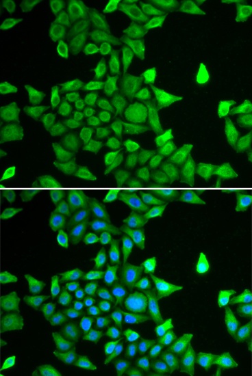
Immunofluorescence analysis of MCF-7 cells using PSMA6 Rabbit pAb (CAB13536). Secondary antibody: Cy3-conjugated Goat anti-Rabbit IgG (H+L) (CABS007) at 1:500 dilution. Blue: DAPI for nuclear staining.