The PSMB7 Antibody (CAB14771) is a high-quality antibody developed for reliable detection and analysis of target proteins. This antibody, produced in rabbits, exhibits high specificity and sensitivity towards human samples, making it ideal for Western blot applications. By binding to PSMB7, researchers can accurately detect and analyze this essential protein in various cell types, facilitating studies in areas such as proteasome function, cell biology, and cancer research.
This antibody is validated for use in WB, IF/ICC, ELISA applications and has demonstrated reactivity against Human, Mouse, Rat samples.
Product Name:
PSMB7 Antibody
SKU:
CAB14771
Size:
20μL, 100μL
Reactivity:
Human, Mouse, Rat
Conjugate:
Unconjugated
Immunogen:
Recombinant protein (or fragment).This information is considered to be commercially sensitive.
Recommended starting concentration is 1 μg/mL. Please optimize the concentration based on your specific assay requirements.
Synonyms:
PSMB7, Z
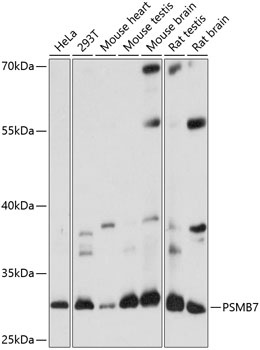
Positive Sample:
HeLa, 293T, Mouse heart, Mouse testis, Mouse brain, Rat testis, Rat brain
Cellular Localization:
Cytoplasm, Nucleus.
Calculated MW:
30kDa
Observed MW:
30kDa
The proteasome is a multicatalytic proteinase complex with a highly ordered ring-shaped 20S core structure. The core structure is composed of 4 rings of 28 non-identical subunits; 2 rings are composed of 7 alpha subunits and 2 rings are composed of 7 beta subunits. Proteasomes are distributed throughout eukaryotic cells at a high concentration and cleave peptides in an ATP/ubiquitin-dependent process in a non-lysosomal pathway. The encoded protein is a member of the proteasome B-type family, also known as the T1B family, and is a 20S core beta subunit in the proteasome. Expression of this catalytic subunit is downregulated by gamma interferon, and proteolytic processing is required to generate a mature subunit. A pseudogene of this gene is located on the long arm of chromosome 14.
Purification Method
Affinity purification
Gene ID
5695
RRID
AB_2761647
Buffer Information
Store at -20℃. Avoid freeze / thaw cycles. Buffer: PBS with 0.01% thimerosal,50% glycerol,pH7.3.
Western blot analysis of various lysates using PSMB7 Rabbit pAb (CAB14771) at 1:1000 dilution. Secondary antibody: HRP-conjugated Goat anti-Rabbit IgG (H+L) (CABS014) at 1:10000 dilution. Lysates/proteins: 25μg per lane. Blocking buffer: 3% nonfat dry milk in TBST. Detection: ECL Basic Kit (AbGn00020). Exposure time: 1s.
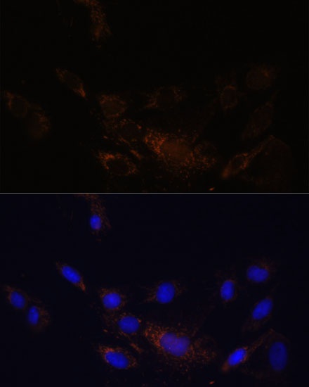
Immunofluorescence analysis of C6 cells using PSMB7 Rabbit pAb (CAB14771) at dilution of 1:100 (40x lens). Secondary antibody: Cy3-conjugated Goat anti-Rabbit IgG (H+L) (CABS007) at 1:500 dilution. Blue: DAPI for nuclear staining.