The PTHLH Antibody (CAB3183) is a high-quality antibody developed for reliable detection and analysis of target proteins. This antibody, produced in rabbits, is highly specific for human samples and has been validated for use in various applications, including Western blot and immunohistochemistry.PTHLH is involved in a wide range of physiological processes, including bone development, cartilage growth, and tooth formation. Dysregulation of PTHLH signaling has been implicated in skeletal disorders, such as osteoporosis, as well as in cancer progression, particularly in breast and prostate cancers.
This antibody is validated for use in WB, IHC-P, ELISA applications and has demonstrated reactivity against Human, Mouse, Rat samples.
Product Name:
PTHLH Antibody
SKU:
CAB3183
Size:
20μL, 100μL
Reactivity:
Human, Mouse, Rat
Conjugate:
Unconjugated
Immunogen:
Recombinant protein (or fragment).This information is considered to be commercially sensitive.
Recommended starting concentration is 1 μg/mL. Please optimize the concentration based on your specific assay requirements.
Synonyms:
HHM, PLP, BDE2, PTHR, PTHRP, PTHLH
Positive Sample:
PC-3, U-251MG
Cellular Localization:
Cytoplasm, Nucleus, Secreted.
Calculated MW:
20kDa
Observed MW:
20kDa
The protein encoded by this gene is a member of the parathyroid hormone family. This hormone, via its receptor, PTHR1, regulates endochondral bone development and epithelial-mesenchymal interactions during the formation of the mammary glands and teeth. It is responsible for most cases of humoral hypercalcemia of malignancy, and mutations in this gene are associated with brachydactyly type E2 (BDE2). Alternatively spliced transcript variants have been found for this gene. There is also evidence for alternative translation initiation from non-AUG (CUG and GUG) start sites, downstream of the initiator AUG codon, resulting in nuclear forms of this hormone.
Purification Method
Affinity purification
Gene ID
5744
RRID
AB_2764970
Buffer Information
Store at -20℃. Avoid freeze / thaw cycles. Buffer: PBS containing 50% glycerol, preserved with proclin300 or sodium azide, pH 7.3.
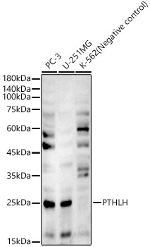
Western blot analysis of various lysates, using PTHLH Rabbit pAb (CAB3183) at 1:8000 dilution. Secondary antibody: HRP-conjugated Goat anti-Rabbit IgG (H+L) (CABS014) at 1:10000 dilution. Lysates/proteins: 25μg per lane. Blocking buffer: 3% nonfat dry milk in TBST. Detection: ECL Basic Kit (AbGn00020). Exposure time: 90s.
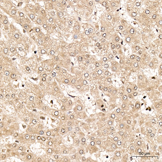
Immunohistochemistry analysis of paraffin-embedded Human liver tissue using PTHLH Rabbit pAb (CAB3183) at a dilution of 1:600 (40x lens). High pressure antigen retrieval performed with 0.01M Citrate Buffer (pH 6.0) prior to IHC staining.
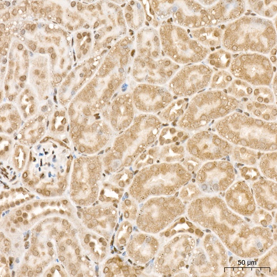
Immunohistochemistry analysis of paraffin-embedded Mouse kidney tissue using PTHLH Rabbit pAb (CAB3183) at a dilution of 1:600 (40x lens). High pressure antigen retrieval performed with 0.01M Citrate Buffer (pH 6.0) prior to IHC staining.
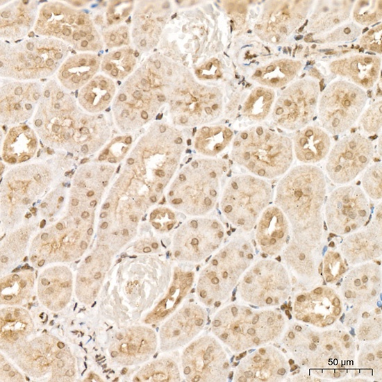
Immunohistochemistry analysis of paraffin-embedded Rat kidney tissue using PTHLH Rabbit pAb (CAB3183) at a dilution of 1:600 (40x lens). High pressure antigen retrieval performed with 0.01M Citrate Buffer (pH 6.0) prior to IHC staining.