The PTX3 Antibody (CAB12669) is a high-quality antibody developed for reliable detection and analysis of target proteins. This antibody, produced in rabbits, is highly specific to PTX3 and has been validated for use in a variety of applications including Western blot and immunohistochemistry.PTX3, also known as pentraxin-related protein PTX3, is a key player in the innate immune response and has been implicated in various inflammatory diseases and conditions.
This antibody is validated for use in WB, ELISA applications and has demonstrated reactivity against Human, Mouse, Rat samples.
Product Name:
PTX3 Antibody
SKU:
CAB12669
Size:
20μL, 100μL
Reactivity:
Human, Mouse, Rat
Conjugate:
Unconjugated
Immunogen:
Recombinant protein (or fragment).This information is considered to be commercially sensitive.
Recommended starting concentration is 1 μg/mL. Please optimize the concentration based on your specific assay requirements.
Synonyms:
TSG-14, TNFAIP5, PTX3
Positive Sample:
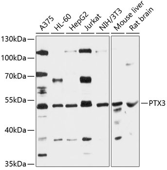
A375, HL-60, HepG2, Jurkat, NIH/3T3, Mouse liver, Rat brain
Cellular Localization:
Secreted.
Calculated MW:
42kDa
Observed MW:
42kDa
This gene encodes a member of the pentraxin protein family. The expression of this protein is induced by inflammatory cytokines in response to inflammatory stimuli in several mesenchymal and epithelial cell types, particularly endothelial cells and mononuclear phagocytes. The protein promotes fibrocyte differentiation and is involved in regulating inflammation and complement activation. It also plays a role in angiogenesis and tissue remodeling. The protein serves as a biomarker for several inflammatory conditions.
Purification Method
Affinity purification
Gene ID
5806
RRID
AB_2759516
Buffer Information
Store at -20℃. Avoid freeze / thaw cycles. Buffer: PBS with 0.01% thimerosal,50% glycerol,pH7.3.
Western blot analysis of various lysates using PTX3 Rabbit pAb (CAB12669) at 1:3000 dilution. Secondary antibody: HRP-conjugated Goat anti-Rabbit IgG (H+L) (CABS014) at 1:10000 dilution. Lysates/proteins: 25μg per lane. Blocking buffer: 3% nonfat dry milk in TBST. Detection: ECL Enhanced Kit (AbGn00021). Exposure time: 30s.