The PUMA Monoclonal Antibody (CAB3752) is a high-quality antibody developed for reliable detection and analysis of target proteins. This antibody, generated from rabbit monoclonal antibodies, is particularly reactive with human samples and has been validated for use in immunohistochemistry and flow cytometry applications.PUMA, also known as p53 upregulated modulator of apoptosis, plays a crucial role in the induction of apoptosis in response to various cellular stresses. Its dysregulation has been implicated in numerous diseases, including cancer and neurodegenerative disorders.
This antibody is validated for use in WB, IHC-P, IF/ICC, ELISA applications and has demonstrated reactivity against Human, Mouse, Rat samples.
Product Name:
PUMA Monoclonal Antibody
SKU:
CAB3752
Size:
20μL, 100μL
Reactivity:
Human, Mouse, Rat
Clone Number:
ARC0247
Conjugate:
Unconjugated
Immunogen:
Synthetic peptide. This information is considered to be commercially sensitive.
This gene encodes a member of the BCL-2 family of proteins. This family member belongs to the BH3-only pro-apoptotic subclass. The protein cooperates with direct activator proteins to induce mitochondrial outer membrane permeabilization and apoptosis. It can bind to anti-apoptotic Bcl-2 family members to induce mitochondrial dysfunction and caspase activation. Because of its pro-apoptotic role, this gene is a potential drug target for cancer therapy and for tissue injury. Alternative splicing results in multiple transcript variants.
Purification Method
Affinity purification
Gene ID
27113
RRID
AB_2863135
Buffer Information
Store at -20℃. Avoid freeze / thaw cycles. Buffer: PBS containing 50% glycerol and 0.05% BSA, preserved with proclin300 or sodium azide, pH 7.3.
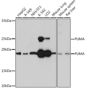
Western blot analysis of various lysates using PUMA Rabbit mAb (CAB3752) at 1:1000 dilution. Secondary antibody: HRP-conjugated Goat anti-Rabbit IgG (H+L) (CABS014) at 1:10000 dilution. Lysates/proteins: 25μg per lane. Blocking buffer: 3% nonfat dry milk in TBST. Detection: ECL Basic Kit (AbGn00020). Exposure time: 30s.
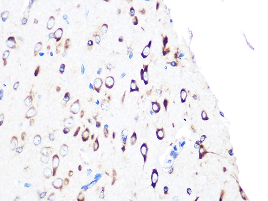
Immunohistochemistry analysis of paraffin-embedded Rat brain using PUMA Rabbit mAb (CAB3752) at dilution of 1:100 (40x lens). Microwave antigen retrieval performed with 0.01M PBS Buffer (pH 7.2) prior to IHC staining.
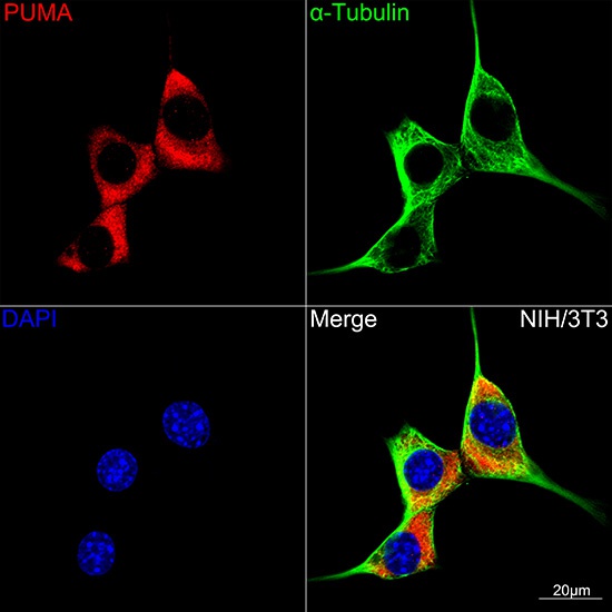
Confocal imaging of NIH/3T3 cells using PUMA Rabbit mAb (CAB3752,dilution 1:100)(Red). The cells were counterstained with α-Tubulin Mouse mAb (AC012,dilution 1:400) (Green). DAPI was used for nuclear staining (blue). Objective: 100x.
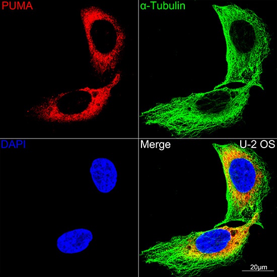
Confocal imaging of U-2 OS cells using PUMA Rabbit mAb (CAB3752,dilution 1:100)(Red). The cells were counterstained with α-Tubulin Mouse mAb (AC012,dilution 1:400) (Green). DAPI was used for nuclear staining (blue). Objective: 100x.