The PVRL4 Antibody (CAB16149) is a high-quality antibody developed for reliable detection and analysis of target proteins. This antibody, generated in rabbits, exhibits high reactivity with human samples and is validated for use in Western blot applications. By targeting the PVRL4 protein, this antibody allows for the detection and analysis of PVRL4 expression in different cell types, making it an ideal choice for studies in cell biology, developmental biology, and cancer research.
This antibody is validated for use in WB, ELISA applications and has demonstrated reactivity against Human, Mouse, Rat samples.
Product Name:
PVRL4 Antibody
SKU:
CAB16149
Size:
20μL, 100μL
Reactivity:
Human, Mouse, Rat
Conjugate:
Unconjugated
Immunogen:
Recombinant protein (or fragment).This information is considered to be commercially sensitive.
Recommended starting concentration is 1 μg/mL. Please optimize the concentration based on your specific assay requirements.
Synonyms:
LNIR, PRR4, EDSS1, PVRL4, nectin-4
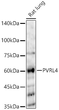
Positive Sample:
Rat lung
Cellular Localization:
Cell Junction, Cell Membrane, Secreted, Single-Pass Type I Membrane Protein, Adherens Junction.
Calculated MW:
55kDa
Observed MW:
62kDa/75kDa
This gene encodes a member of the nectin family. The encoded protein contains two immunoglobulin-like (Ig-like) C2-type domains and one Ig-like V-type domain. It is involved in cell adhesion through trans-homophilic and -heterophilic interactions. It is a single-pass type I membrane protein. The soluble form is produced by proteolytic cleavage at the cell surface by the metalloproteinase ADAM17/TACE. The secreted form is found in both breast tumor cell lines and breast tumor patients. Mutations in this gene are the cause of ectodermal dysplasia-syndactyly syndrome type 1, an autosomal recessive disorder. Alternatively spliced transcript variants have been found but the full-length nature of the variant has not been determined.
Purification Method
Affinity purification
Gene ID
81607
RRID
AB_2763596
Buffer Information
Store at -20℃. Avoid freeze / thaw cycles. Buffer: PBS containing 50% glycerol, preserved with proclin300 or sodium azide, pH 7.3.
Western blot analysis of various lysates, using PVRL4 Rabbit pAb (CAB16149) at 1:2000 dilution. Secondary antibody: HRP-conjugated Goat anti-Rabbit IgG (H+L) (CABS014) at 1:10000 dilution. Lysates/proteins: 25μg per lane. Blocking buffer: 3% nonfat dry milk in TBST. Detection: ECL Basic Kit (AbGn00020). Exposure time: 90s.