The ASC/TMS1 Antibody (CAB16672) is a high-quality antibody developed for reliable detection and analysis of target proteins. This gene encodes an adaptor protein that is composed of two protein-protein interaction domains: a N-terminal PYRIN-PAAD-DAPIN domain (PYD) and a C-terminal caspase-recruitment domain (CARD). The PYD and CARD domains are members of the six-helix bundle death domain-fold superfamily that mediates assembly of large signaling complexes in the inflammatory and apoptotic signaling pathways via the activation of caspase. In normal cells, this protein is localized to the cytoplasm; however, in cells undergoing apoptosis, it forms ball-like aggregates near the nuclear periphery. Two transcript variants encoding different isoforms have been found for this gene.
This antibody is validated for use in WB, IHC-P, IF/ICC, IP, ELISA applications and has demonstrated reactivity against Human, Mouse, Rat samples.
Product Name:
ASC/TMS1 Antibody
SKU:
CAB16672
Size:
100μL, 20μL
Reactivity:
Human, Mouse, Rat
Conjugate:
Unconjugated
Immunogen:
Recombinant protein (or fragment).This information is considered to be commercially sensitive.
Tested Applications:
WBIHC-PIF/ICCIPELISA
Recommended Dilution:
WB
1:1000 - 1:6000
IHC-P
1:50 - 1:200
IF/ICC
1:50 - 1:200
IP
0.5μg-4μg antibody for 400μg-600μg extracts of whole cells
ELISA
Recommended starting concentration is 1 μg/mL. Please optimize the concentration based on your specific assay requirements.
This gene encodes an adaptor protein that is composed of two protein-protein interaction domains: a N-terminal PYRIN-PAAD-DAPIN domain (PYD) and a C-terminal caspase-recruitment domain (CARD). The PYD and CARD domains are members of the six-helix bundle death domain-fold superfamily that mediates assembly of large signaling complexes in the inflammatory and apoptotic signaling pathways via the activation of caspase. In normal cells, this protein is localized to the cytoplasm; however, in cells undergoing apoptosis, it forms ball-like aggregates near the nuclear periphery. Two transcript variants encoding different isoforms have been found for this gene.
Purification Method
Affinity purification
Gene ID
29108
RRID
AB_2771891
Buffer Information
Store at -20℃. Avoid freeze / thaw cycles. Buffer: PBS containing 50% glycerol, preserved with proclin300 or sodium azide, pH 7.3.
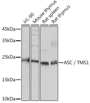
Western blot analysis of various lysates using ASC/TMS1 Rabbit pAb (CAB16672) at 1:3000 dilution. Secondary antibody: HRP-conjugated Goat anti-Rabbit IgG (H+L) (AS014) at 1:10000 dilution. Lysates/proteins: 25μg per lane. Blocking buffer: 3% nonfat dry milk in TBST. Detection: ECL Basic Kit (AbGn00020). Exposure time: 10s.
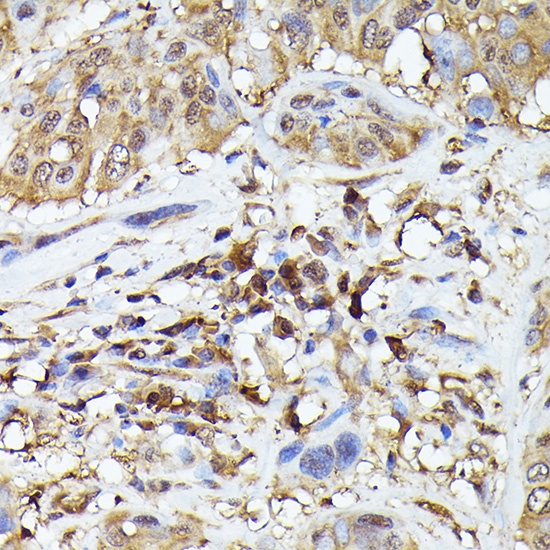
Immunohistochemistry analysis of paraffin-embedded Human esophageal cancer using ASC/TMS1 Rabbit pAb (CAB16672) at dilution of 1:200 (40x lens). High pressure antigen retrieval performed with 0.01M Citrate buffer (pH 6.0) prior to IHC staining.
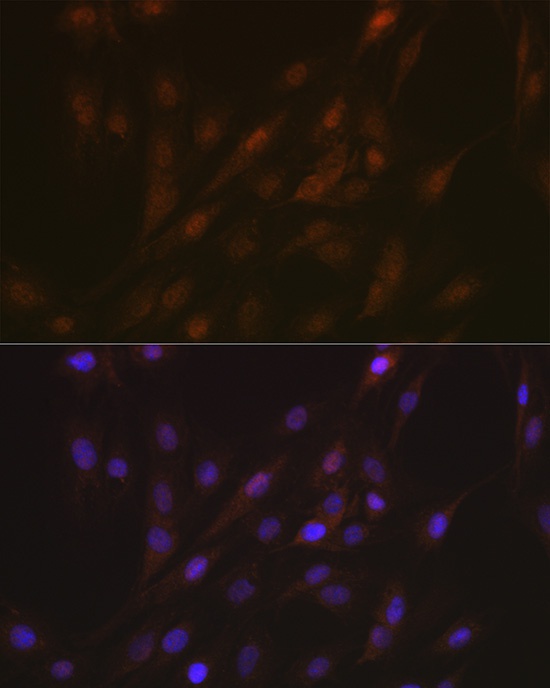
Immunofluorescence analysis of C6 cells using ASC/TMS1 Rabbit pAb (CAB16672) at dilution of 1:100 (40x lens). Secondary antibody: Cy3-conjugated Goat anti-Rabbit IgG (H+L) (AS007) at 1:500 dilution. Blue: DAPI for nuclear staining.
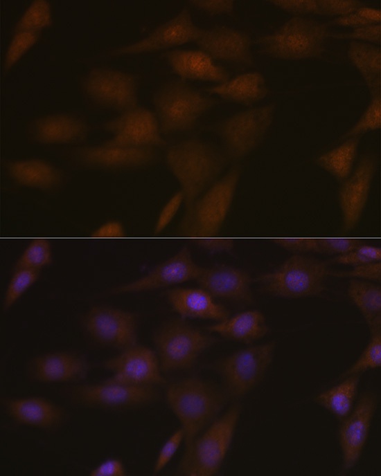
Immunofluorescence analysis of NIH-3T3 cells using ASC/TMS1 Rabbit pAb (CAB16672) at dilution of 1:100 (40x lens). Secondary antibody: Cy3-conjugated Goat anti-Rabbit IgG (H+L) (AS007) at 1:500 dilution. Blue: DAPI for nuclear staining.
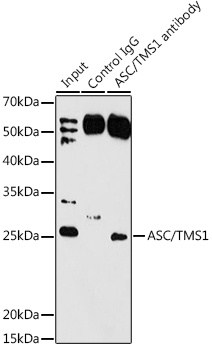
Immunoprecipitation analysis of 200 μg extracts of Rat lung using 3 μg ASC/TMS1 antibody (CAB16672). Western blot was performed from the immunoprecipitate using ASC/TMS1 antibody (CAB16672) at a dilution of 1:1000.


 ELISA Kit (HUFI03337)")
 ELISA Kit (HUFI03087)")