The PYGB Antibody (CAB6402) is a high-quality antibody developed for reliable detection and analysis of target proteins. This antibody, produced in rabbits, has high reactivity with human samples and has been validated for use in Western blotting applications. By specifically binding to PYGB, researchers can easily detect and analyze the enzyme in various cell types, making it an ideal choice for studies in metabolism, diabetes, and other metabolic disorders.PYGB is an essential enzyme involved in breaking down glycogen, the main storage form of glucose in the body.
This antibody is validated for use in WB, IF/ICC, IP, ELISA applications and has demonstrated reactivity against Human, Mouse, Rat samples.
Product Name:
PYGB Antibody
SKU:
CAB6402
Size:
20μL, 100μL
Reactivity:
Human, Mouse, Rat
Conjugate:
Unconjugated
Immunogen:
Recombinant protein (or fragment).This information is considered to be commercially sensitive.
The protein encoded by this gene is a glycogen phosphorylase found predominantly in the brain. The encoded protein forms homodimers which can associate into homotetramers, the enzymatically active form of glycogen phosphorylase. The activity of this enzyme is positively regulated by AMP and negatively regulated by ATP, ADP, and glucose-6-phosphate. This enzyme catalyzes the rate-determining step in glycogen degradation.
Purification Method
Affinity purification
Gene ID
5834
RRID
AB_2767004
Buffer Information
Store at -20℃. Avoid freeze / thaw cycles. Buffer: PBS containing 50% glycerol, preserved with proclin300 or sodium azide, pH 7.3.
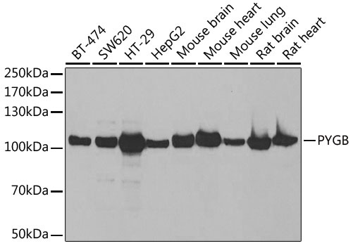
Western blot analysis of various lysates using PYGB Rabbit pAb (CAB6402) at 1:1000 dilution. Secondary antibody: HRP-conjugated Goat anti-Rabbit IgG (H+L) (CABS014) at 1:10000 dilution. Lysates/proteins: 25μg per lane. Blocking buffer: 3% nonfat dry milk in TBST. Detection: ECL Basic Kit (AbGn00020). Exposure time: 30s.
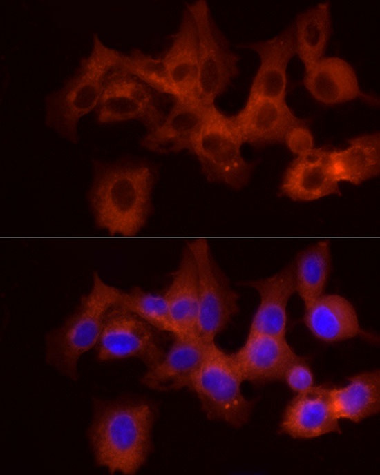
Immunofluorescence analysis of HepG2 cells using PYGB Rabbit pAb (CAB6402) at dilution of 1:100 (40x lens). Secondary antibody: Cy3-conjugated Goat anti-Rabbit IgG (H+L) (CABS007) at 1:500 dilution. Blue: DAPI for nuclear staining.
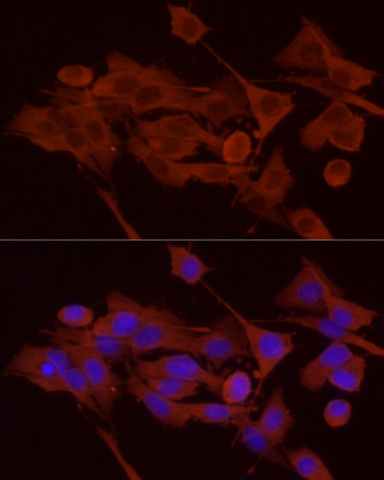
Immunofluorescence analysis of PC-12 cells using PYGB Rabbit pAb (CAB6402) at dilution of 1:100 (40x lens). Secondary antibody: Cy3-conjugated Goat anti-Rabbit IgG (H+L) (CABS007) at 1:500 dilution. Blue: DAPI for nuclear staining.
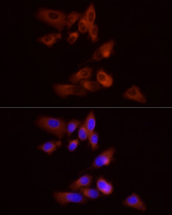
Immunofluorescence analysis of PC-3 cells using PYGB Rabbit pAb (CAB6402) at dilution of 1:100 (40x lens). Secondary antibody: Cy3-conjugated Goat anti-Rabbit IgG (H+L) (CABS007) at 1:500 dilution. Blue: DAPI for nuclear staining.