The RSPO1 Antibody (CAB8289) is a high-quality antibody developed for reliable detection and analysis of target proteins. This gene encodes a secreted activator protein with two cysteine-rich, furin-like domains and one thrombospondin type 1 domain. The encoded protein is a ligand for leucine-rich repeat-containing G-protein coupled receptors (LGR proteins) and positively regulates the Wnt signaling pathway. In mice, the protein induces the rapid onset of crypt cell proliferation and increases intestinal epithelial healing, providing a protective effect against chemotherapy-induced adverse effects. Alternative splicing results in multiple transcript variants.
This antibody is validated for use in WB, IHC-P, IF/ICC, ELISA applications and has demonstrated reactivity against Human, Mouse, Rat samples.
Product Name:
RSPO1 Antibody
SKU:
CAB8289
Size:
100μL, 20μL
Reactivity:
Human, Mouse, Rat
Conjugate:
Unconjugated
Immunogen:
Recombinant protein (or fragment).This information is considered to be commercially sensitive.
Tested Applications:
WBIHC-PIF/ICCELISA
Recommended Dilution:
WB
1:500 - 1:1000
IHC-P
1:50 - 1:200
IF/ICC
1:50 - 1:200
ELISA
Recommended starting concentration is 1 μg/mL. Please optimize the concentration based on your specific assay requirements.
Synonyms:
RSPO, CRISTIN3, RSPO1
Positive Sample:
SKOV3, Rat pancreas
Cellular Localization:
Secreted.
Calculated MW:
29kDa
Observed MW:
29kDa
This gene encodes a secreted activator protein with two cysteine-rich, furin-like domains and one thrombospondin type 1 domain. The encoded protein is a ligand for leucine-rich repeat-containing G-protein coupled receptors (LGR proteins) and positively regulates the Wnt signaling pathway. In mice, the protein induces the rapid onset of crypt cell proliferation and increases intestinal epithelial healing, providing a protective effect against chemotherapy-induced adverse effects. Alternative splicing results in multiple transcript variants.
Purification Method
Affinity purification
Gene ID
284654
RRID
AB_2772123
Buffer Information
Store at -20℃. Avoid freeze / thaw cycles. Buffer: PBS containing 50% glycerol, preserved with proclin300 or sodium azide, pH 7.3.
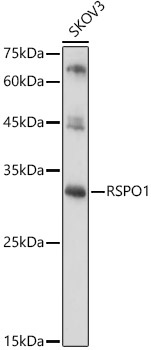
Western blot analysis of lysates from SKOV3 cells, using RSPO1 Rabbit pAb (CAB8289) at 1:1000 dilution. Secondary antibody: HRP-conjugated Goat anti-Rabbit IgG (H+L) (AS014) at 1:10000 dilution. Lysates/proteins: 25μg per lane. Blocking buffer: 3% nonfat dry milk in TBST. Detection: ECL Basic Kit (AbGn00020). Exposure time: 60s.
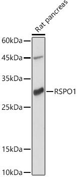
Western blot analysis of lysates from Rat pancreas, using RSPO1 Rabbit pAb (CAB8289) at 1:1000 dilution. Secondary antibody: HRP-conjugated Goat anti-Rabbit IgG (H+L) (AS014) at 1:10000 dilution. Lysates/proteins: 25μg per lane. Blocking buffer: 3% nonfat dry milk in TBST. Detection: ECL Basic Kit (AbGn00020). Exposure time: 180s.
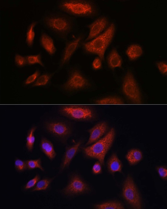
Immunofluorescence analysis of A-549 cells using RSPO1 Rabbit pAb (CAB8289) at dilution of 1:100 (40x lens). Secondary antibody: Cy3-conjugated Goat anti-Rabbit IgG (H+L) (AS007) at 1:500 dilution. Blue: DAPI for nuclear staining.
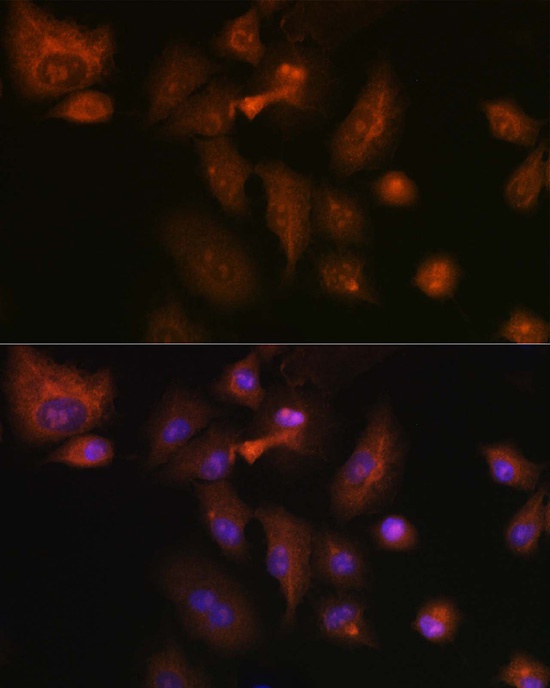
Immunofluorescence analysis of A-549 cells using RSPO1 Rabbit pAb (CAB8289) at dilution of 1:100 (40x lens). Secondary antibody: Cy3-conjugated Goat anti-Rabbit IgG (H+L) (AS007) at 1:500 dilution. Blue: DAPI for nuclear staining.