The RAB11FIP5 Antibody (CAB18142) is a high-quality antibody developed for reliable detection and analysis of target proteins. This rabbit polyclonal antibody is highly specific for human samples and has been validated for use in various applications, including Western blot and immunofluorescence.Rab11FIP5, also known as Rab11 family-interacting protein 5, plays a crucial role in the recycling of endocytic vesicles and the regulation of membrane trafficking pathways. Its involvement in these processes makes it a key player in cell physiology and is implicated in various cellular processes, including cell migration, cytokinesis, and cell adhesion.
This antibody is validated for use in WB, IF/ICC, ELISA applications and has demonstrated reactivity against Human, Mouse, Rat samples.
Product Name:
RAB11FIP5 Antibody
SKU:
CAB18142
Size:
20μL, 100μL
Reactivity:
Human, Mouse, Rat
Conjugate:
Unconjugated
Immunogen:
Recombinant protein (or fragment).This information is considered to be commercially sensitive.
Enables gamma-tubulin binding activity. Involved in cellular response to acidic pH; negative regulation of adiponectin secretion; and regulation of protein localization to cell surface. Located in centriolar satellite and mitochondrial outer membrane.
Purification Method
Affinity purification
Gene ID
26056
RRID
AB_2861933
Buffer Information
Store at -20℃. Avoid freeze / thaw cycles. Buffer: PBS containing 50% glycerol, preserved with proclin300 or sodium azide, pH 7.3.
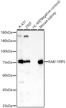
Western blot analysis of various lysates, using RAB11FIP5 Rabbit pAb (CAB18142) at 1:800 dilution. Secondary antibody: HRP-conjugated Goat anti-Rabbit IgG (H+L) (CABS014) at 1:10000 dilution. Lysates/proteins: 25μg per lane. Blocking buffer: 3% nonfat dry milk in TBST. Detection: ECL Basic Kit (AbGn00020). Exposure time: 60s.
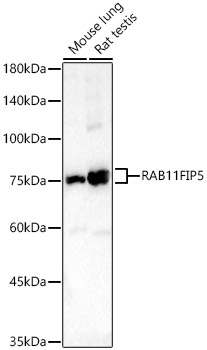
Western blot analysis of various lysates, using RAB11FIP5 Rabbit pAb (CAB18142) at 1:800 dilution. Secondary antibody: HRP-conjugated Goat anti-Rabbit IgG (H+L) (CABS014) at 1:10000 dilution. Lysates/proteins: 25μg per lane. Blocking buffer: 3% nonfat dry milk in TBST. Detection: ECL Basic Kit (AbGn00020). Exposure time: 180s.
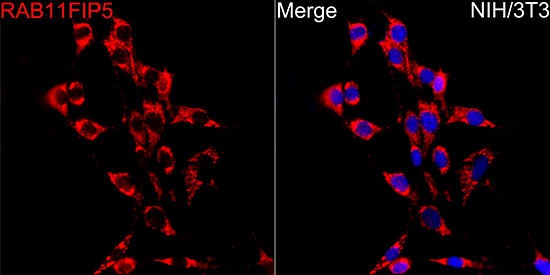
Immunofluorescence analysis of NIH/3T3 cells using RAB11FIP5 Rabbit pAb (CAB18142) at dilution of 1:50 (40x lens). Secondary antibody: Cy3-conjugated Goat anti-Rabbit IgG (H+L) (CABS007) at 1:500 dilution. Blue: DAPI for nuclear staining.