The Rad50 Antibody (CAB3078) is a high-quality antibody developed for reliable detection and analysis of target proteins. Raised in rabbits, this antibody is highly specific for human RAD50 samples and has been validated for use in Western blot applications.RAD50 is a member of the MRN complex, which is essential for detecting and repairing DNA double-strand breaks. Dysregulation of RAD50 has been implicated in various diseases, including cancer and genetic disorders.
This antibody is validated for use in WB, IHC-P, IP, ELISA applications and has demonstrated reactivity against Human, Mouse, Rat samples.
Product Name:
Rad50 Antibody
SKU:
CAB3078
Size:
20μL, 100μL
Reactivity:
Human, Mouse, Rat
Conjugate:
Unconjugated
Immunogen:
Synthetic peptide. This information is considered to be commercially sensitive.
0.5μg-4μg antibody for 200μg-400μg extracts of whole cells
ELISA
Recommended starting concentration is 1 μg/mL. Please optimize the concentration based on your specific assay requirements.
Synonyms:
NBSLD, RAD502, hRad50, Rad50
Positive Sample:
HeLa, K-562, U-937, Mouse testis, Mouse thymus, Rat testis
Cellular Localization:
Chromosome, Nucleus, Telomere.
Calculated MW:
154kDa
Observed MW:
154kDa
The protein encoded by this gene is highly similar to Saccharomyces cerevisiae Rad50, a protein involved in DNA double-strand break repair. This protein forms a complex with MRE11 and NBS1. The protein complex binds to DNA and displays numerous enzymatic activities that are required for nonhomologous joining of DNA ends. This protein, cooperating with its partners, is important for DNA double-strand break repair, cell cycle checkpoint activation, telomere maintenance, and meiotic recombination. Knockout studies of the mouse homolog suggest this gene is essential for cell growth and viability. Mutations in this gene are the cause of Nijmegen breakage syndrome-like disorder.
Purification Method
Affinity purification
Gene ID
10111
RRID
AB_2764881
Buffer Information
Store at -20℃. Avoid freeze / thaw cycles. Buffer: PBS containing 50% glycerol, preserved with proclin300 or sodium azide, pH 7.3.
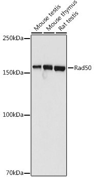
Western blot analysis of various lysates using Rad50 Rabbit pAb (CAB3078) at 1:1000 dilution. Secondary antibody: HRP-conjugated Goat anti-Rabbit IgG (H+L) (CABS014) at 1:10000 dilution. Lysates/proteins: 25μg per lane. Blocking buffer: 3% nonfat dry milk in TBST. Detection: ECL Basic Kit (AbGn00020). Exposure time: 3s.
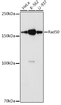
Western blot analysis of various lysates using Rad50 Rabbit pAb (CAB3078) at 1:1000 dilution. Secondary antibody: HRP-conjugated Goat anti-Rabbit IgG (H+L) (CABS014) at 1:10000 dilution. Lysates/proteins: 25μg per lane. Blocking buffer: 3% nonfat dry milk in TBST. Detection: ECL Basic Kit (AbGn00020). Exposure time: 90s.
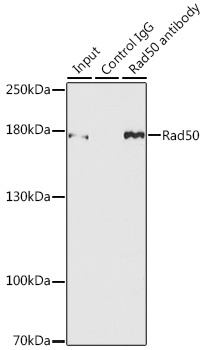
Immunoprecipitation analysis of 150 μg extracts of MCF7 cells using 3 μg Rad50 antibody (CAB3078). Western blot was performed from the immunoprecipitate using Rad50 antibody (CAB3078) at a dilution of 1:500.