The Recombinant Human CSF-3/G-CSF Protein is a biologically active recombinant protein that plays a significant role in various cellular processes and signaling pathways in human biology. This protein is widely employed in immunological research, cell biology studies, protein-protein interaction analyses, and therapeutic development, providing researchers with a reliable tool for investigating CSF-3/G-CSF function and its implications in health and disease.
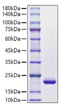
This product (SKU: RPCB1230) is produced using HEK293 cells and features a C-6His tag for convenient detection and purification. The protein exhibits a calculated molecular weight of 22.82 kDa with an observed molecular weight of 20 kDa under denaturing conditions, achieving ≥ 95 % as determined by SDS-PAGE;≥ 95 % as determined by HPLC.. Functional bioactivity has been validated through rigorous quality control assays, confirming its suitability for demanding research applications.
High quality, high purity and low endotoxin recombinant Recombinant Human CSF-3/G-CSF Protein (RPCB1230), tested reactivity in HEK293 cells and has been validated in SDS-PAGE.100% guaranteed.
Endotoxin:
< 0.1 EU/μg of the protein by LAL method.
Purity:
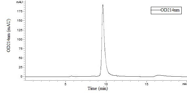
≥ 95 % as determined by SDS-PAGE;≥ 95 % as determined by HPLC.
Formulation:
Lyophilized from a 0.22 μm filtered solution of PBS, pH 7.4.
Bio-Activity:
Measured in a cell proliferation assay using NFS-60 mouse myelogenous leukemia lymphoblast cells. The ED 50 for this effect is 41-164 pg/mL.
Reconstitution:
Centrifuge the vial before opening. Reconstitute to a concentration of 0.1-0.5 mg/mL in sterile distilled water. Avoid vortex or vigorously pipetting the protein. For long term storage, it is recommended to add a carrier protein or stablizer (e.g. 0.1% BSA, 5% HSA, 10% FBS or 5% Trehalose), and aliquot the reconstituted protein solution to minimize free-thaw cycles.
Storage:
Store at -20℃.Store the lyophilized protein at -20℃ to -80 ℃ up to 1 year from the date of receipt. After reconstitution, the protein solution is stable at -20℃ for 3 months, at 2-8℃ for up to 1 week.
Granulocyte-colony stimulating factor (G-CSF) is a growth factor and an essential cytokine belonging to the CSF family of hormone-like glycoproteins. It is produced by numerous cell types including immune and endothelial cells. G-CSF binding to its receptor G-CSF-R which belongs to the cytokine receptor type I family depends on the interaction of alpha-helical motifs of the former and two fibronectin type III as well as an immunoglobulin-like domain of the latter. Recent animal studies have also revealed that G-CSF activates multiple signaling pathways, such as Akt and also the Janus family kinase-2 and signal transducer and activation of transcription-3 (Jak2-STAT3) pathway, thereby promoting survival, proliferation, differentiation and mobilisation of haematopoietic stem and progenitor cells. G-CSF is a cytokine that have been demonstrated to improve cardiac function and perfusion in myocardial infarction. And it was initially evaluated as a stem cell mobilizer and erythropoietin as a cytoprotective agent. G-CSF prevents left ventricular remodeling after myocardial infarction by decreasing cardiomyocyte death and by increasing the number of blood vessels, suggesting the importance of direct actions of G-CSF on the myocardium rather than through mobilization and differentiation of stem cells. Accordingly, recombinant human (rh)G-CSF has been extensively used in clinical haematology and oncology to enable bone marrow transplantation or to treat chemotherapy-associated neutropenia. In preclinical study, G-CSF improved cardiac function and perfusion by angiomyogenesis and protection of cardiomyocytes in myocardial infarction.
Recombinant Human CSF-3/G-CSF Protein was determined by SDS-PAGE under reducing conditions with Coomassie Blue.
Recombinant Human CSF-3/G-CSF Protein is greater than 95% as determined by SEC-HPLC.
")
")
")
")
")

 (RPES8023)")