The REP5S1 Polyclonal Antibody (PAC058432) is a valuable tool for researchers studying the Replication Pericentriolar Material 5 (REP5) protein. This antibody, produced in rabbits, exhibits high reactivity towards human samples and has been validated for use in Western blot applications. By binding specifically to the REP5 protein, this antibody enables accurate detection and analysis in a variety of cell types, making it suitable for investigations in molecular biology and cancer research.The REP5 protein is a key player in cellular replication processes, particularly in the pericentriolar region of the cell.
Its function is essential for maintaining genomic stability and proper cell division, highlighting its relevance in understanding cancer development and progression. By studying the role of REP5, researchers can gain insights into potential therapeutic targets for cancer treatment and other diseases involving aberrant cell replication.Overall, the REP5S1 Polyclonal Antibody offers researchers a valuable tool for investigating the functions of the REP5 protein in various biological processes, paving the way for advancements in cancer research and molecular biology studies.
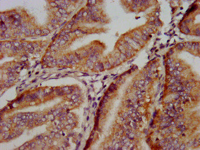
IHC image of PACO58432 diluted at 1:500 and staining in paraffin-embedded human endometrial cancer performed on a Leica BondTM system. After dewaxing and hydration, antigen retrieval was mediated by high pressure in a citrate buffer (pH 6.0). Section was blocked with 10% normal goat serum 30min at RT. Then primary antibody (1% BSA) was incubated at 4°C overnight. The primary is detected by a biotinylated secondary antibody and visualized using an HRP conjugated SP system.
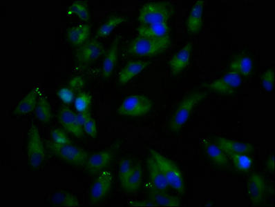
Immunofluorescence staining of Hela cells with PACO58432 at 1:166, counter-stained with DAPI. The cells were fixed in 4% formaldehyde, permeabilized using 0.2% Triton X-100 and blocked in 10% normal Goat Serum. The cells were then incubated with the antibody overnight at 4°C. The secondary antibody was Alexa Fluor 488-congugated AffiniPure Goat Anti-Rabbit IgG(H+L).
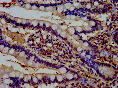
IHC image of PACO58432 diluted at 1:500 and staining in paraffin-embedded human small intestine tissue performed on a Leica BondTM system. After dewaxing and hydration, antigen retrieval was mediated by high pressure in a citrate buffer (pH 6.0). Section was blocked with 10% normal goat serum 30min at RT. Then primary antibody (1% BSA) was incubated at 4°C overnight. The primary is detected by a biotinylated secondary antibody and visualized using an HRP conjugated SP system.
Background:
May coordinate the cellular actions of activated EGF receptors and Ral-GTPases.
Synonyms:
RalBP1-associated Eps domain-containing protein 1 (RalBP1-interacting protein 1), REPS1
UniProt Protein Function:
May coordinate the cellular actions of activated EGF receptors and Ral-GTPases.
UniProt Protein Details:
NCBI Summary:
This gene encodes a signaling adaptor protein with two EH domains that interacts with proteins that participate in signaling, endocytosis and cytoskeletal changes. The encoded protein has been found in association with intersectin 1 and Src homology 3-domain growth factor receptor-bound 2-like (endophilin) interacting protein 1 when intersectin 1 was isolated from clathrin-coated pits. The encoded protein has also been shown to interact with amphiphysin, a cytoplasmic protein at the surface of synaptic vesicles. Alternative splicing results in multiple transcript variants encoding different isoforms. [provided by RefSeq, Mar 2014]


 ELISA Kit (HUFI03057)")

