The RGR Antibody (CAB7925) is a high-quality antibody developed for reliable detection and analysis of target proteins. This antibody, produced in rabbits, exhibits high reactivity with human samples and has been validated for use in Western blot applications. By binding to the RGR protein, this antibody allows for precise detection and analysis in different cell types, making it an essential component for studies in various fields such as neuroscience, vision research, and retinal degeneration.
This antibody is validated for use in WB, ELISA applications and has demonstrated reactivity against Rat samples.
Product Name:
RGR Antibody
SKU:
CAB7925
Size:
20μL, 100μL
Reactivity:
Rat
Conjugate:
Unconjugated
Immunogen:
Recombinant protein (or fragment).This information is considered to be commercially sensitive.
Recommended starting concentration is 1 μg/mL. Please optimize the concentration based on your specific assay requirements.
Synonyms:
RP44, RGR
Positive Sample:
rat spinal cord
Cellular Localization:
Membrane, Multi-Pass Membrane Protein.
Calculated MW:
32kDa
Observed MW:
32kDa
This gene encodes a putative retinal G-protein coupled receptor. The gene is a member of the opsin subfamily of the 7 transmembrane, G-protein coupled receptor 1 family. Like other opsins which bind retinaldehyde, it contains a conserved lysine residue in the seventh transmembrane domain. The protein acts as a photoisomerase to catalyze the conversion of all-trans-retinal to 11-cis-retinal. The reverse isomerization occurs with rhodopsin in retinal photoreceptor cells. The protein is exclusively expressed in tissue adjacent to retinal photoreceptor cells, the retinal pigment epithelium and Mueller cells. This gene may be associated with autosomal recessive and autosomal dominant retinitis pigmentosa (arRP and adRP, respectively). Alternative splicing results in multiple transcript variants encoding different isoforms.
Purification Method
Affinity purification
Gene ID
5995
RRID
AB_2771996
Buffer Information
Store at -20℃. Avoid freeze / thaw cycles. Buffer: PBS containing 50% glycerol, preserved with proclin300 or sodium azide, pH 7.3.
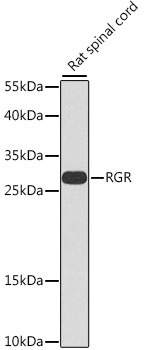
Western blot analysis of lysates from rat spinal cord, using RGR Rabbit pAb (CAB7925) at 1:1000 dilution. Secondary antibody: HRP-conjugated Goat anti-Rabbit IgG (H+L) (CABS014) at 1:10000 dilution. Lysates/proteins: 25μg per lane. Blocking buffer: 3% nonfat dry milk in TBST. Detection: ECL Basic Kit (AbGn00020). Exposure time: 90s.