The RHOH Antibody (CAB6356) is a high-quality antibody developed for reliable detection and analysis of target proteins. The protein encoded by this gene is a member of the Ras superfamily of guanosine triphosphate (GTP)-metabolizing enzymes. The encoded protein is expressed in hematopoietic cells, where it functions as a negative regulator of cell growth and survival. This gene may be hypermutated or misexpressed in leukemias and lymphomas. Chromosomal translocations in non-Hodgkin's lymphoma occur between this locus and B-cell CLL/lymphoma 6 (BCL6) on chromosome 3, leading to the production of fusion transcripts. Alternative splicing in the 5' untranslated region results in multiple transcript variants that encode the same protein.
This antibody is validated for use in WB, IF/ICC, ELISA applications and has demonstrated reactivity against Human, Mouse samples.
Product Name:
RHOH Antibody
SKU:
CAB6356
Size:
100μL, 20μL
Reactivity:
Human, Mouse
Conjugate:
Unconjugated
Immunogen:
Recombinant protein (or fragment).This information is considered to be commercially sensitive.
Tested Applications:
WBIF/ICCELISA
Recommended Dilution:
WB
1:500 - 1:2000
IF/ICC
1:10 - 1:100
ELISA
Recommended starting concentration is 1 μg/mL. Please optimize the concentration based on your specific assay requirements.
The protein encoded by this gene is a member of the Ras superfamily of guanosine triphosphate (GTP)-metabolizing enzymes. The encoded protein is expressed in hematopoietic cells, where it functions as a negative regulator of cell growth and survival. This gene may be hypermutated or misexpressed in leukemias and lymphomas. Chromosomal translocations in non-Hodgkin's lymphoma occur between this locus and B-cell CLL/lymphoma 6 (BCL6) on chromosome 3, leading to the production of fusion transcripts. Alternative splicing in the 5' untranslated region results in multiple transcript variants that encode the same protein.
Purification Method
Affinity purification
Gene ID
399
RRID
AB_2766958
Buffer Information
Store at -20℃. Avoid freeze / thaw cycles. Buffer: PBS containing 50% glycerol, preserved with proclin300 or sodium azide, pH 7.3.
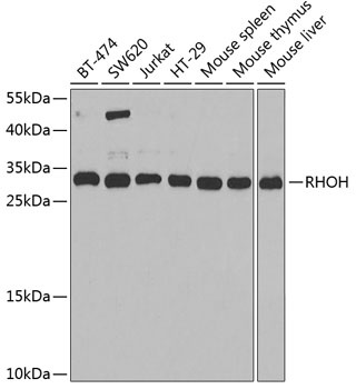
Western blot analysis of various lysates using RHOH Rabbit pAb (CAB6356) at 1:1000 dilution. Secondary antibody: HRP-conjugated Goat anti-Rabbit IgG (H+L) (AS014) at 1:10000 dilution. Lysates/proteins: 25μg per lane. Blocking buffer: 3% nonfat dry milk in TBST. Detection: ECL Basic Kit (AbGn00020). Exposure time: 90s.
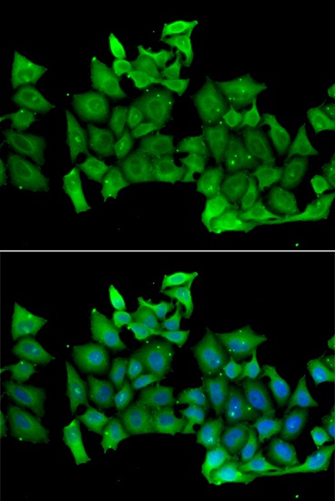
Immunofluorescence analysis of MCF-7 cells using RHOH Rabbit pAb (CAB6356). Secondary antibody: Cy3-conjugated Goat anti-Rabbit IgG (H+L) (AS007) at 1:500 dilution. Blue: DAPI for nuclear staining.