The RMND5A Antibody (CAB14924) is a high-quality antibody developed for reliable detection and analysis of target proteins. This antibody, produced in rabbits, is highly specific to human samples and has been validated for use in Western blot applications. By targeting the RMND5A protein, this antibody allows for the detection and analysis of RMND5A in various cell types, making it ideal for studies in genetics, cell biology, and cancer research.RMND5A, also known as Required for meiotic nuclear division protein 5A, plays a crucial role in maintaining genome stability and proper cell division. Its function in DNA replication suggests its importance in understanding cell proliferation and potential implications for cancer development.
This antibody is validated for use in WB, ELISA applications and has demonstrated reactivity against Human, Mouse, Rat samples.
Product Name:
RMND5A Antibody
SKU:
CAB14924
Size:
20μL, 100μL
Reactivity:
Human, Mouse, Rat
Conjugate:
Unconjugated
Immunogen:
Recombinant protein (or fragment).This information is considered to be commercially sensitive.
Recommended starting concentration is 1 μg/mL. Please optimize the concentration based on your specific assay requirements.
Synonyms:
CTLH, GID2, RMD5, GID2A, p44CTLH, RMND5A
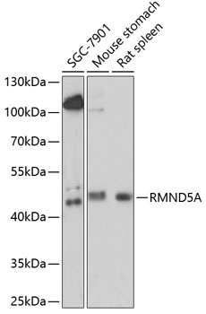
Positive Sample:
SGC-7901, Mouse stomach, Rat spleen
Cellular Localization:
Cytoplasm, Nucleoplasm, Nucleus.
Calculated MW:
44kDa
Observed MW:
44kDa
Predicted to enable metal ion binding activity and ubiquitin protein ligase activity. Predicted to contribute to ubiquitin-protein transferase activity. Predicted to be involved in proteasome-mediated ubiquitin-dependent protein catabolic process and protein polyubiquitination. Located in cytoplasm and nucleoplasm. Part of ubiquitin ligase complex.
Purification Method
Affinity purification
Gene ID
64795
RRID
AB_2761806
Buffer Information
Store at -20℃. Avoid freeze / thaw cycles. Buffer: PBS with 0.01% thimerosal,50% glycerol,pH7.3.
Western blot analysis of various lysates using RMND5A Rabbit pAb (CAB14924) at 1:1000 dilution. Secondary antibody: HRP-conjugated Goat anti-Rabbit IgG (H+L) (CABS014) at 1:10000 dilution. Lysates/proteins: 25μg per lane. Blocking buffer: 3% nonfat dry milk in TBST. Detection: ECL Basic Kit (AbGn00020). Exposure time: 90s.