The ROR1 Antibody (CAB3315) is a high-quality antibody developed for reliable detection and analysis of target proteins. This antibody, generated in rabbits, is highly specific and reactive with human samples, making it ideal for use in Western blot applications.ROR1 is a cell surface protein that is implicated in various cellular processes, including cell proliferation, survival, and migration. It plays a role in cancer development and has been identified as a potential therapeutic target in certain types of cancer.By targeting the ROR1 protein, researchers can investigate its role in cancer progression and potentially develop targeted therapies for ROR1-expressing cancers.
This antibody is validated for use in WB, ELISA applications and has demonstrated reactivity against Rat samples.
Product Name:
ROR1 Antibody
SKU:
CAB3315
Size:
20μL, 100μL
Reactivity:
Rat
Conjugate:
Unconjugated
Immunogen:
Recombinant protein (or fragment).This information is considered to be commercially sensitive.
Recommended starting concentration is 1 μg/mL. Please optimize the concentration based on your specific assay requirements.
Synonyms:
NTRKR1, dJ537F10.1, ROR1
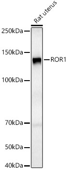
Positive Sample:
Rat uterus
Cellular Localization:
Membrane, Single-Pass Type I Membrane Protein.
Calculated MW:
104kDa
Observed MW:
135kDa
This gene encodes a receptor tyrosine kinase-like orphan receptor that modulates neurite growth in the central nervous system. The encoded protein is a glycosylated type I membrane protein that belongs to the ROR subfamily of cell surface receptors. It is a pseudokinase that lacks catalytic activity and may interact with the non-canonical Wnt signalling pathway. This gene is highly expressed during early embryonic development but expressed at very low levels in adult tissues. Increased expression of this gene is associated with B-cell chronic lymphocytic leukaemia. Alternative splicing results in multiple transcript variants encoding different isoforms.
Purification Method
Affinity purification
Gene ID
4919
RRID
AB_2765045
Buffer Information
Store at -20℃. Avoid freeze / thaw cycles. Buffer: PBS containing 50% glycerol, preserved with proclin300 or sodium azide, pH 7.3.
Western blot analysis of lysates from Rat uterus, using ROR1 Rabbit pAb (CAB3315) at 1:800 dilution. Secondary antibody: HRP-conjugated Goat anti-Rabbit IgG (H+L) (CABS014) at 1:10000 dilution. Lysates/proteins: 25μg per lane. Blocking buffer: 3% nonfat dry milk in TBST. Detection: ECL Basic Kit (AbGn00020). Exposure time: 90s.