The RPL10A Antibody (CAB5925) is a high-quality antibody developed for reliable detection and analysis of target proteins. The antibody, raised in rabbits, is highly specific and reactive with human, mouse, and rat samples, making it versatile for use in various applications such as Western blot and immunohistochemistry.RPL10A is a key player in ribosome assembly and function, essential for the translation of genetic information into proteins. Its involvement in cellular processes makes it a valuable target for research in molecular biology, cancer biology, and developmental biology.
This antibody is validated for use in WB, IHC-P, IF/ICC, ELISA applications and has demonstrated reactivity against Human, Mouse, Rat samples.
Product Name:
RPL10A Antibody
SKU:
CAB5925
Size:
20μL, 100μL
Reactivity:
Human, Mouse, Rat
Conjugate:
Unconjugated
Immunogen:
Recombinant protein (or fragment).This information is considered to be commercially sensitive.
Ribosomes, the organelles that catalyze protein synthesis, consist of a small 40S subunit and a large 60S subunit. Together these subunits are composed of 4 RNA species and approximately 80 structurally distinct proteins. This gene encodes a ribosomal protein that is a component of the 60S subunit. The protein belongs to the L1P family of ribosomal proteins. It is located in the cytoplasm. The expression of this gene is downregulated in the thymus by cyclosporin-A (CsA), an immunosuppressive drug. Studies in mice have shown that the expression of the ribosomal protein L10a gene is downregulated in neural precursor cells during development. This gene previously was referred to as NEDD6 (neural precursor cell expressed, developmentally downregulated 6), but it has been renamed RPL10A (ribosomal protein 10a). As is typical for genes encoding ribosomal proteins, there are multiple processed pseudogenes of this gene dispersed through the genome.
Purification Method
Affinity purification
Gene ID
4736
RRID
AB_2766664
Buffer Information
Store at -20℃. Avoid freeze / thaw cycles. Buffer: PBS containing 50% glycerol, preserved with proclin300 or sodium azide, pH 7.3.
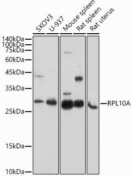
Western blot analysis of various lysates using RPL10A Rabbit pAb (CAB5925) at 1:1000 dilution. Secondary antibody: HRP-conjugated Goat anti-Rabbit IgG (H+L) (CABS014) at 1:10000 dilution. Lysates/proteins: 25μg per lane. Blocking buffer: 3% nonfat dry milk in TBST. Detection: ECL Enhanced Kit (AbGn00021). Exposure time: 180s.
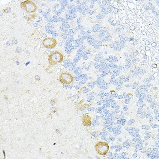
Immunohistochemistry analysis of paraffin-embedded Rat cerebellum using RPL10A Rabbit pAb (CAB5925) at dilution of 1:100 (40x lens). High pressure antigen retrieval performed with 0.01M Citrate buffer (pH 6.0) prior to IHC staining.
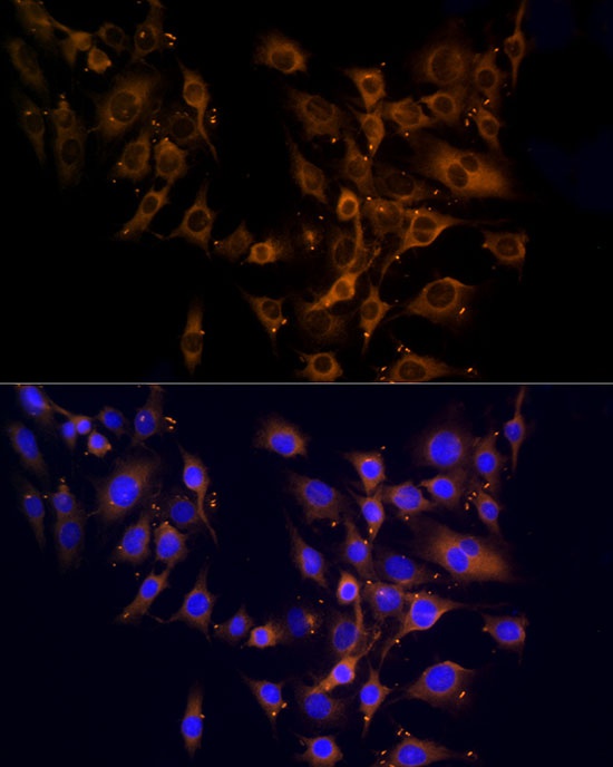
Immunofluorescence analysis of C6 cells using RPL10A Rabbit pAb (CAB5925) at dilution of 1:100 (40x lens). Secondary antibody: Cy3-conjugated Goat anti-Rabbit IgG (H+L) (CABS007) at 1:500 dilution. Blue: DAPI for nuclear staining.