The SATB1 Antibody (CAB5800) is a high-quality antibody developed for reliable detection and analysis of target proteins. This antibody, produced in rabbits, is highly specific for human samples and has been validated for use in Western blotting and immunohistochemistry applications.SATB1, or Special AT-rich Sequence-Binding Protein 1, plays a vital role in the organization of chromatin structure and the regulation of gene expression. Its involvement in various cellular processes, including cell differentiation, proliferation, and immune response, makes it a valuable target for research in immunology, cancer, and developmental biology.
This antibody is validated for use in WB, IHC-P, IP, ELISA, IF-P applications and has demonstrated reactivity against Human, Mouse, Rat samples.
Product Name:
SATB1 Antibody
SKU:
CAB5800
Size:
20μL, 100μL
Reactivity:
Human, Mouse, Rat
Conjugate:
Unconjugated
Immunogen:
Recombinant protein (or fragment).This information is considered to be commercially sensitive.
0.5μg-4μg antibody for 200μg-400μg extracts of whole cells
IF-P
1:50 - 1:200
IHC-P
1:50 - 1:200
ELISA
Recommended starting concentration is 1 μg/mL. Please optimize the concentration based on your specific assay requirements.
Synonyms:
DEFDA, KTZSL, SATB1
Positive Sample:
Jurkat
Cellular Localization:
Nucleus, Nucleus Matrix, Pml Body.
Calculated MW:
86kDa
Observed MW:
100kDa
This gene encodes a matrix protein which binds nuclear matrix and scaffold-associating DNAs through a unique nuclear architecture. The protein recruits chromatin-remodeling factors in order to regulate chromatin structure and gene expression.
Purification Method
Affinity purification
Gene ID
6304
RRID
AB_2766552
Buffer Information
Store at -20℃. Avoid freeze / thaw cycles. Buffer: PBS containing 50% glycerol, preserved with proclin300 or sodium azide, pH 7.3.
Western blot analysis of lysates from Jurkat cells, using SATB1 Rabbit pAb (CAB5800) at 1:1000 dilution. Secondary antibody: HRP-conjugated Goat anti-Rabbit IgG (H+L) (CABS014) at 1:10000 dilution. Lysates/proteins: 25μg per lane. Blocking buffer: 3% nonfat dry milk in TBST. Detection: ECL Basic Kit (AbGn00020). Exposure time: 1s.
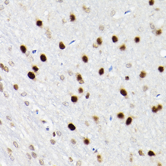
Immunohistochemistry analysis of paraffin-embedded Rat brain using SATB1 Rabbit pAb (CAB5800) at dilution of 1:200 (40x lens). High pressure antigen retrieval performed with 0.01M Citrate buffer (pH 6.0) prior to IHC staining.
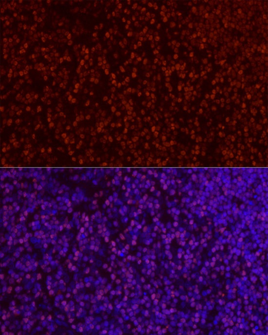
Immunofluorescence analysis of paraffin-embedded rat thymus using SATB1 Rabbit pAb (CAB5800) at dilution of 1:50 (40x lens). Secondary antibody: Cy3-conjugated Goat anti-Rabbit IgG (H+L) (CABS007) at 1:500 dilution. Blue: DAPI for nuclear staining.
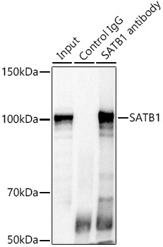
Immunoprecipitation analysis of 300 μg extracts of Jurkat cells using 3 μg SATB1 antibody (CAB5800). Western blot was performed from the immunoprecipitate using SATB1 antibody (CAB5800) at a dilution of 1:500.