The SAV1 Antibody (CAB9980) is a high-quality antibody developed for reliable detection and analysis of target proteins. This antibody, produced in rabbits, exhibits high specificity and sensitivity towards human samples, making it an ideal choice for Western blot applications. By binding to the SAV1 protein, this antibody enables precise detection and analysis in a variety of cell types, facilitating investigations in developmental biology and cancer research.SAV1, also known as Salvador homolog 1, plays a crucial role in controlling organ size and tissue regeneration by modulating cell proliferation and apoptosis.
This antibody is validated for use in WB, ELISA applications and has demonstrated reactivity against Human, Mouse samples.
Product Name:
SAV1 Antibody
SKU:
CAB9980
Size:
20μL, 100μL
Reactivity:
Human, Mouse
Conjugate:
Unconjugated
Immunogen:
Recombinant protein (or fragment).This information is considered to be commercially sensitive.
Recommended starting concentration is 1 μg/mL. Please optimize the concentration based on your specific assay requirements.
Synonyms:
SAV, WW45, WWP4, SAV1
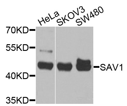
Positive Sample:
HeLa, SKOV3, SW480
Cellular Localization:
Cytoplasm, Nucleus.
Calculated MW:
45kDa
Observed MW:
45kDa
WW domain-containing proteins are found in all eukaryotes and play an important role in the regulation of a wide variety of cellular functions such as protein degradation, transcription, and RNA splicing. This gene encodes a protein with two WW domains, a SARAH domain, and a coiled-coil region and is ubiquitously expressed in adult tissues. This protein binds to MST1 (mammalian sterile 20-like kinase 1) and promotes MST1-induced apoptosis. It has also been shown to bind to HAX1 (hematopoietic cell-specific protein 1 (HS1)-associated protein X-1) and to attenuate the anti-apoptotic effects of HAX1. Studies in human and mouse suggest this gene acts as a tumor suppressor.
Purification Method
Affinity purification
Gene ID
60485
RRID
AB_2772146
Buffer Information
Store at -20℃. Avoid freeze / thaw cycles. Buffer: PBS with 0.09% Sodium azide,50% glycerol,pH7.3.
Western blot analysis of various lysates using SAV1 Rabbit pAb (CAB9980) at 1:1000 dilution. Secondary antibody: HRP-conjugated Goat anti-Rabbit IgG (H+L) (CABS014) at 1:10000 dilution. Lysates/proteins: 25μg per lane. Blocking buffer: 3% nonfat dry milk in TBST. Detection: ECL Basic Kit (AbGn00020). Exposure time: 30s.