The SDHA Monoclonal Antibody (CAB13852) is a high-quality antibody developed for reliable detection and analysis of target proteins. This antibody is raised in rabbits and is highly reactive with human samples, making it a valuable tool for studying mitochondrial function and metabolism.Validated for use in Western blot applications, this antibody specifically binds to the SDHA protein, allowing for easy detection and analysis in various cell types. Its reliability and specificity make it ideal for research in biochemistry, cell biology, and metabolism.
This antibody is validated for use in WB, IF/ICC, ELISA, IF-P applications and has demonstrated reactivity against Human, Mouse, Rat samples.
Product Name:
SDHA Monoclonal Antibody
SKU:
CAB13852
Size:
20μL, 100μL
Reactivity:
Human, Mouse, Rat
Clone Number:
ARC0726
Conjugate:
Unconjugated
Immunogen:
Synthetic peptide. This information is considered to be commercially sensitive.
This gene encodes a major catalytic subunit of succinate-ubiquinone oxidoreductase, a complex of the mitochondrial respiratory chain. The complex is composed of four nuclear-encoded subunits and is localized in the mitochondrial inner membrane. Mutations in this gene have been associated with a form of mitochondrial respiratory chain deficiency known as Leigh Syndrome. A pseudogene has been identified on chromosome 3q29. Alternatively spliced transcript variants encoding different isoforms have been found for this gene.
Purification Method
Affinity purification
Gene ID
6389
RRID
AB_2861697
Buffer Information
Store at -20℃. Avoid freeze / thaw cycles. Buffer: PBS containing 50% glycerol and 0.05% BSA, preserved with proclin300 or sodium azide, pH 7.3.
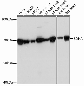
Western blot analysis of various lysates using SDHA Rabbit mAb (CAB13852) at 1:1000 dilution incubated overnight at 4℃. Secondary antibody: HRP-conjugated Goat anti-Rabbit IgG (H+L) (CABS014) at 1:10000 dilution. Lysates/proteins: 25 μg per lane. Blocking buffer: 3% nonfat dry milk in TBST. Detection: ECL Basic Kit (AbGn00020). Exposure time: 1 s.
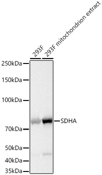
Western blot analysis of lysates from 293F cells using SDHA Rabbit mAb (CAB13852) at 1:1000 dilution incubated overnight at 4℃. Mitochondrion extracts derived from 293F cells. Secondary antibody: HRP-conjugated Goat anti-Rabbit IgG (H+L) (CABS014) at 1:10000 dilution. Lysates/proteins: 25 μg per lane. Blocking buffer: 3% nonfat dry milk in TBST. Detection: ECL Basic Kit (AbGn00020). Exposure time: 1 s.
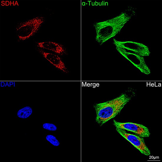
Confocal imaging of HeLa cells using SDHA Rabbit mAb (CAB13852, dilution 1:200) followed by a further incubation with Cy3-conjugated Goat anti-Rabbit IgG (H+L) (CABS007, dilution 1:500) (Red). The cells were counterstained with α-Tubulin Mouse mAb (AC012, dilution 1:400) followed by incubation with ABflo® 488-conjugated Goat Anti-Mouse IgG (H+L) Ab (CABS076, dilution 1:500) (Green). DAPI was used for nuclear staining (Blue). Objective: 100x.