The SEL1L Antibody (CAB12073) is a high-quality antibody developed for reliable detection and analysis of target proteins. This antibody, generated in rabbits, exhibits high reactivity with human samples and has been validated for use in Western blot applications. By specifically binding to the SEL1L protein, this antibody allows for precise detection and analysis in a variety of cell types, making it an essential reagent for investigations in the fields of cell biology and protein degradation pathways.
This antibody is validated for use in WB, IF/ICC, ELISA applications and has demonstrated reactivity against Human, Mouse, Rat samples.
Product Name:
SEL1L Antibody
SKU:
CAB12073
Size:
20μL, 100μL
Reactivity:
Human, Mouse, Rat
Conjugate:
Unconjugated
Immunogen:
Recombinant protein (or fragment).This information is considered to be commercially sensitive.
Recommended starting concentration is 1 μg/mL. Please optimize the concentration based on your specific assay requirements.
Synonyms:
Hrd3, SEL1L1, PRO1063, SEL1-LIKE, SEL1L
Positive Sample:
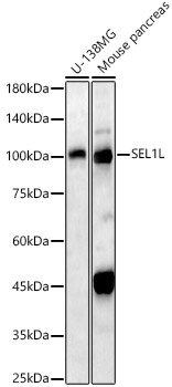
U-138MG, Mouse pancreas
Cellular Localization:
Endoplasmic Reticulum Membrane, Single-Pass Type I Membrane Protein.
Calculated MW:
89kDa
Observed MW:
100kDa
The protein encoded by this gene is part of a protein complex required for the retrotranslocation or dislocation of misfolded proteins from the endoplasmic reticulum lumen to the cytosol, where they are degraded by the proteasome in a ubiquitin-dependent manner. Alternatively spliced transcript variants encoding different isoforms have been found for this gene.
Purification Method
Affinity purification
Gene ID
6400
RRID
AB_2758977
Buffer Information
Store at -20℃. Avoid freeze / thaw cycles. Buffer: PBS containing 50% glycerol, preserved with proclin300 or sodium azide, pH 7.3.
Western blot analysis of various lysates, using SEL1L Rabbit pAb (CAB12073) at 1:1000 dilution. Secondary antibody: HRP-conjugated Goat anti-Rabbit IgG (H+L) (CABS014) at 1:10000 dilution. Lysates/proteins: 25μg per lane. Blocking buffer: 3% nonfat dry milk in TBST. Detection: ECL Basic Kit (AbGn00020). Exposure time: 180s.
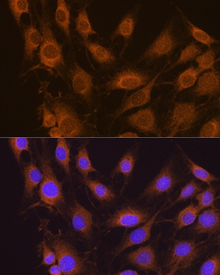
Immunofluorescence analysis of C6 cells using SEL1L Rabbit pAb (CAB12073) at dilution of 1:100. Blue: DAPI for nuclear staining.