The SEMA4A Antibody (CAB17205) is a high-quality antibody developed for reliable detection and analysis of target proteins. This antibody, produced in rabbits, demonstrates high reactivity with human samples and is validated for use in Western blot applications. By targeting the SEMA4A protein, this antibody enables the detection and analysis of SEMA4A expression in a variety of cell types, making it an essential tool for studies in immunology, neurobiology, and cancer research.
This antibody is validated for use in WB, IF/ICC, ELISA applications and has demonstrated reactivity against Human, Mouse, Rat samples.
Product Name:
SEMA4A Antibody
SKU:
CAB17205
Size:
20μL, 100μL
Reactivity:
Human, Mouse, Rat
Immunogen:
Recombinant protein (or fragment).This information is considered to be commercially sensitive.
Recommended starting concentration is 1 μg/mL. Please optimize the concentration based on your specific assay requirements.
Synonyms:
RP35, SEMB, SEMAB, CORD10, SEMA4A
Positive Sample:
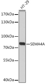
HT-29
Cellular Localization:
Extracellular Space, Plasma Membrane.
Calculated MW:
84kDa
Observed MW:
83kDa
This gene encodes a member of the semaphorin family of soluble and transmembrane proteins. Semaphorins are involved in numerous functions, including axon guidance, morphogenesis, carcinogenesis, and immunomodulation. The encoded protein is a single-pass type I membrane protein containing an immunoglobulin-like C2-type domain, a PSI domain and a sema domain. It inhibits axonal extension by providing local signals to specify territories inaccessible for growing axons. It is an activator of T-cell-mediated immunity and suppresses vascular endothelial growth factor (VEGF)-mediated endothelial cell migration and proliferation in vitro and angiogenesis in vivo. Mutations in this gene are associated with retinal degenerative diseases including retinitis pigmentosa type 35 (RP35) and cone-rod dystrophy type 10 (CORD10). Multiple alternatively spliced transcript variants encoding different isoforms have been identified.
Purification Method
Affinity purification
Gene ID
64218
RRID
AB_2772175
Buffer Information
Store at -20℃. Avoid freeze / thaw cycles. Buffer: PBS with 0.01% thimerosal,50% glycerol,pH7.3.
Western blot analysis of lysates from HT-29 cells, using SEMA4A Rabbit pAb (CAB17205) at 1:3000 dilution. Secondary antibody: HRP-conjugated Goat anti-Rabbit IgG (H+L) (CABS014) at 1:10000 dilution. Lysates/proteins: 25μg per lane. Blocking buffer: 3% nonfat dry milk in TBST. Detection: ECL Basic Kit (AbGn00020). Exposure time: 90s.
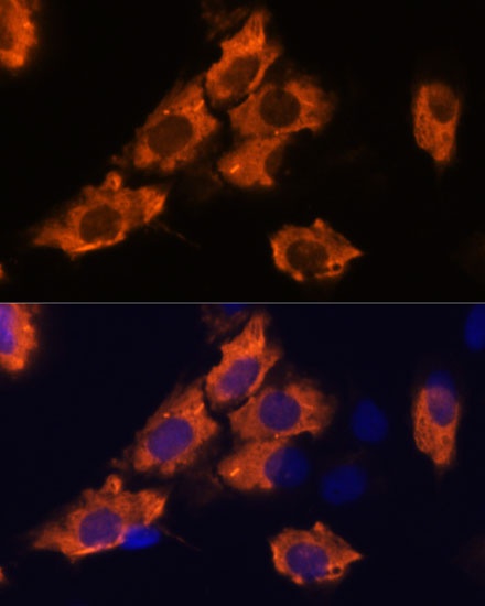
Immunofluorescence analysis of C6 cells using SEMA4A Rabbit pAb (CAB17205) at dilution of 1:100. Secondary antibody: Cy3-conjugated Goat anti-Rabbit IgG (H+L) (CABS007) at 1:500 dilution. Blue: DAPI for nuclear staining.