The Septin 2 Antibody (CAB5801) is a high-quality antibody developed for reliable detection and analysis of target proteins. Raised in rabbits, this antibody is highly specific to human samples and is validated for use in Western blot applications.Septin 2 is known for its involvement in the formation of cellular structures such as the cytoskeleton and cell division machinery.
This antibody is validated for use in WB, IF/ICC, ELISA applications and has demonstrated reactivity against Human, Mouse samples.
Product Name:
Septin 2 Antibody
SKU:
CAB5801
Size:
20μL, 100μL
Reactivity:
Human, Mouse
Conjugate:
Unconjugated
Immunogen:
Recombinant protein (or fragment).This information is considered to be commercially sensitive.
Enables identical protein binding activity. Predicted to be involved in several processes, including cilium assembly; regulation of exocytosis; and smoothened signaling pathway. Predicted to act upstream of or within regulation of L-glutamate import across plasma membrane and regulation of protein localization. Located in several cellular components, including cytoskeleton; photoreceptor connecting cilium; and sperm annulus. Part of septin complex.
Purification Method
Affinity purification
Gene ID
4735
RRID
AB_2766553
Buffer Information
Store at -20℃. Avoid freeze / thaw cycles. Buffer: PBS containing 50% glycerol, preserved with proclin300 or sodium azide, pH 7.3.
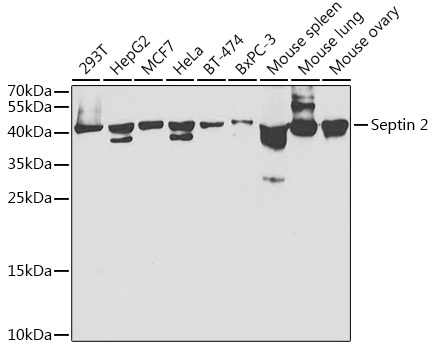
Western blot analysis of various lysates using Septin 2 Rabbit pAb (CAB5801) at 1:1000 dilution. Secondary antibody: HRP-conjugated Goat anti-Rabbit IgG (H+L) (CABS014) at 1:10000 dilution. Lysates/proteins: 25μg per lane. Blocking buffer: 3% nonfat dry milk in TBST. Detection: ECL Basic Kit (AbGn00020). Exposure time: 30s.
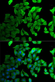
Immunofluorescence analysis of U2OS cells using Septin 2 Rabbit pAb (CAB5801). Secondary antibody: Cy3-conjugated Goat anti-Rabbit IgG (H+L) (CABS007) at 1:500 dilution. Blue: DAPI for nuclear staining.