The Septin 5 Antibody (CAB12953) is a high-quality antibody developed for reliable detection and analysis of target proteins. This gene is a member of the septin gene family of nucleotide binding proteins, originally described in yeast as cell division cycle regulatory proteins. Septins are highly conserved in yeast, Drosophila, and mouse and appear to regulate cytoskeletal organization. Disruption of septin function disturbs cytokinesis and results in large multinucleate or polyploid cells. This gene is mapped to 22q11, the region frequently deleted in DiGeorge and velocardiofacial syndromes. A translocation involving the MLL gene and this gene has also been reported in patients with acute myeloid leukemia. Alternative splicing results in multiple transcript variants. The presence of a non-consensus polyA signal (AACAAT) in this gene also results in read-through transcription into the downstream neighboring gene (GP1BB; platelet glycoprotein Ib), whereby larger, non-coding transcripts are produced. RRID AB_2759799 Gene ID 5413 Swiss Prot Synonym H5; SEPT5; CDCREL; PNUTL1; CDCREL1; CDCREL-1; HCDCREL-1; Septin 5
This antibody is validated for use in WB, IHC-P, IF/ICC, ELISA applications and has demonstrated reactivity against Human, Mouse, Rat samples.
Product Name:
Septin 5 Antibody
SKU:
CAB12953
Size:
100μL, 20μL
Reactivity:
Human, Mouse, Rat
Clone Number:
-
Conjugate:
Unconjugated
Immunogen:
Recombinant protein (or fragment).This information is considered to be commercially sensitive.
Tested Applications:
WBIHC-PIF/ICCELISA
Recommended Dilution:
WB
1:1000 - 1:5000
IHC-P
1:100 - 1:200
IF
/
ICC
1:50 - 1:200
ELISA
Recommended starting concentration is 1 μg/mL. Please optimize the concentration based on your specific assay requirements.
This gene is a member of the septin gene family of nucleotide binding proteins, originally described in yeast as cell division cycle regulatory proteins. Septins are highly conserved in yeast, Drosophila, and mouse and appear to regulate cytoskeletal organization. Disruption of septin function disturbs cytokinesis and results in large multinucleate or polyploid cells. This gene is mapped to 22q11, the region frequently deleted in DiGeorge and velocardiofacial syndromes. A translocation involving the MLL gene and this gene has also been reported in patients with acute myeloid leukemia. Alternative splicing results in multiple transcript variants. The presence of a non-consensus polyA signal (AACAAT) in this gene also results in read-through transcription into the downstream neighboring gene (GP1BB; platelet glycoprotein Ib), whereby larger, non-coding transcripts are produced. RRID AB_2759799 Gene ID 5413 Swiss Prot Synonym H5; SEPT5; CDCREL; PNUTL1; CDCREL1; CDCREL-1; HCDCREL-1; Septin 5
Purification Method:
Affinity purification
Gene ID:
5413
RRID:
AB_2759799
Buffer Information:
Store at -20℃. Avoid freeze / thaw cycles. Buffer: PBS with 0.01% thimerosal,50% glycerol,pH7.3.
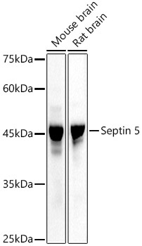
Western blot analysis of various lysates, using Septin 5 Rabbit pAb (CAB12953) at 1:3000 dilution. Secondary antibody: HRP-conjugated Goat anti-Rabbit IgG (H+L) (AS014) at 1:10000 dilution. Lysates/proteins: 25μg per lane. Blocking buffer: 3% nonfat dry milk in TBST. Detection: ECL Basic Kit (AbGn00020). Exposure time: 10s.
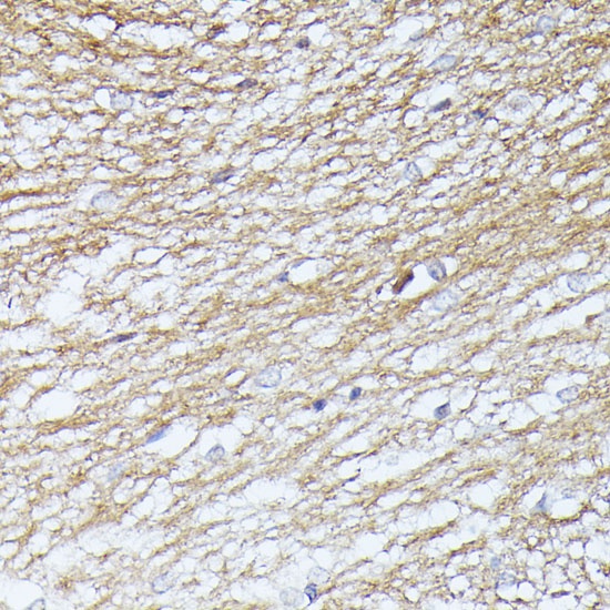
Immunohistochemistry analysis of paraffin-embedded Rat brain using Septin 5 Rabbit pAb (CAB12953) at dilution of 1:100 (40x lens). Microwave antigen retrieval performed with 0.01M PBS Buffer (pH 7.2) prior to IHC staining.
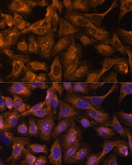
Immunofluorescence analysis of U-2 OS cells using Septin 5 Rabbit pAb (CAB12953) at dilution of 1:100. Secondary antibody: Cy3-conjugated Goat anti-Rabbit IgG (H+L) (AS007) at 1:500 dilution. Blue: DAPI for nuclear staining.