The SERCA2/ATP2A2 Monoclonal Antibody (CAB11692) is a high-quality antibody developed for reliable detection and analysis of target proteins. This monoclonal antibody, produced in rabbits, is highly specific and reactive with human samples, making it a reliable tool for various research applications.SERCA2 plays a vital role in muscle contraction and relaxation by regulating calcium levels within the cell. Dysregulation of SERCA2 has been linked to various diseases, including heart failure, muscular dystrophy, and neurodegenerative disorders. By targeting SERCA2 with this monoclonal antibody, researchers can study its expression, localization, and function in different cell types and tissues.
This antibody is validated for use in WB, IF/ICC, IP, ELISA applications and has demonstrated reactivity against Human, Mouse, Rat samples.
Product Name:
SERCA2/ATP2A2 Monoclonal Antibody
SKU:
CAB11692
Size:
20μL, 100μL
Reactivity:
Human, Mouse, Rat
Clone Number:
ARC0679
Conjugate:
Unconjugated
Immunogen:
Synthetic peptide. This information is considered to be commercially sensitive.
This gene encodes one of the SERCA Ca(2+)-ATPases, which are intracellular pumps located in the sarcoplasmic or endoplasmic reticula of the skeletal muscle. This enzyme catalyzes the hydrolysis of ATP coupled with the translocation of calcium from the cytosol into the sarcoplasmic reticulum lumen, and is involved in regulation of the contraction/relaxation cycle. Mutations in this gene cause Darier-White disease, also known as keratosis follicularis, an autosomal dominant skin disorder characterized by loss of adhesion between epidermal cells and abnormal keratinization. Other types of mutations in this gene have been associated with various forms of muscular dystrophies. Alternative splicing results in multiple transcript variants encoding different isoforms.
Purification Method
Affinity purification
Gene ID
488
RRID
AB_2861632
Buffer Information
Store at -20℃. Avoid freeze / thaw cycles. Buffer: PBS containing 50% glycerol and 0.05% BSA, preserved with proclin300 or sodium azide, pH 7.3.
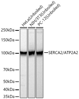
Western blot analysis of various lysates using SERCA2/ATP2A2 Rabbit mAb (CAB11692) at 1:10000 dilution incubated at room temperature for 1.5 hours. Secondary antibody: HRP-conjugated Goat anti-Rabbit IgG (H+L) (CABS014) at 1:10000 dilution. Lysates/proteins: 25 μg per lane. Blocking buffer: 3% nonfat dry milk in TBST. Detection: ECL Basic Kit (AbGn00020). Exposure time: 60 s.
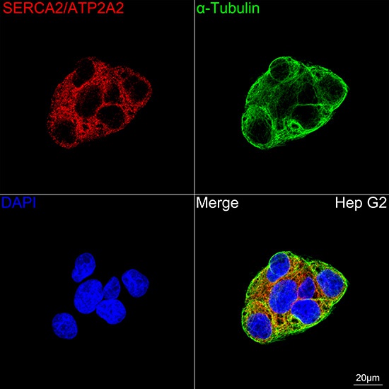
Confocal imaging of Hep G2 cells using SERCA2/ATP2A2 Rabbit mAb (CAB11692, dilution 1:100) followed by a further incubation with Cy3 Goat Anti-Rabbit IgG (H+L) (CABS007, dilution 1:500) (Red). The cells were counterstained with α-Tubulin Mouse mAb (AC012, dilution 1:400) followed by incubation with ABflo® 488-conjugated Goat Anti-Mouse IgG (H+L) Ab (CABS076, dilution 1:500) (Green). DAPI was used for nuclear staining (Blue). Objective: 100x.