The SF3A3 Antibody (CAB4465) is a high-quality antibody developed for reliable detection and analysis of target proteins. This antibody, produced in rabbits, is highly specific to human samples and is validated for use in Western blotting applications. By targeting the SF3A3 protein, this antibody allows for the detection and analysis of SF3A3 in various cell types, making it ideal for studies in molecular biology and gene expression regulation.SF3A3 plays a crucial role in the splicing process, ensuring the accurate removal of introns from pre-mRNA molecules.
This antibody is validated for use in WB, IHC-P, IF/ICC, ELISA applications and has demonstrated reactivity against Human, Mouse, Rat samples.
Product Name:
SF3A3 Antibody
SKU:
CAB4465
Size:
20μL, 100μL
Reactivity:
Human, Mouse, Rat
Conjugate:
Unconjugated
Immunogen:
Recombinant protein (or fragment).This information is considered to be commercially sensitive.
Recommended starting concentration is 1 μg/mL. Please optimize the concentration based on your specific assay requirements.
Synonyms:
PRP9, PRPF9, SAP61, SF3a60, SF3A3
Positive Sample:
293T, NIH/3T3, Raji, Mouse heart, Rat lung
Cellular Localization:
Nucleus Speckle.
Calculated MW:
59kDa
Observed MW:
59kDa
This gene encodes subunit 3 of the splicing factor 3a protein complex. The splicing factor 3a heterotrimer includes subunits 1, 2 and 3 and is necessary for the in vitro conversion of 15S U2 snRNP into an active 17S particle that performs pre-mRNA splicing. Subunit 3 interacts with subunit 1 through its amino-terminus while the zinc finger domain of subunit 3 plays a role in its binding to the 15S U2 snRNP. This gene has a pseudogene on chromosome 20. Alternative splicing results in multiple transcript variants.
Purification Method
Affinity purification
Gene ID
10946
RRID
AB_2614926
Buffer Information
Store at -20℃. Avoid freeze / thaw cycles. Buffer: PBS containing 50% glycerol, preserved with proclin300 or sodium azide, pH 7.3.
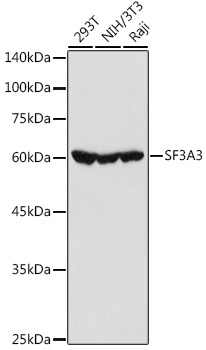
Western blot analysis of various lysates using SF3A3 Rabbit pAb (CAB4465) at 1:1000 dilution. Secondary antibody: HRP-conjugated Goat anti-Rabbit IgG (H+L) (CABS014) at 1:10000 dilution. Lysates/proteins: 25μg per lane. Blocking buffer: 3% nonfat dry milk in TBST. Detection: ECL Basic Kit (AbGn00020). Exposure time: 10s.
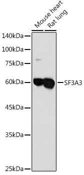
Western blot analysis of various lysates using SF3A3 Rabbit pAb (CAB4465) at 1:1000 dilution. Secondary antibody: HRP-conjugated Goat anti-Rabbit IgG (H+L) (CABS014) at 1:10000 dilution. Lysates/proteins: 25μg per lane. Blocking buffer: 3% nonfat dry milk in TBST. Detection: ECL Basic Kit (AbGn00020). Exposure time: 30s.
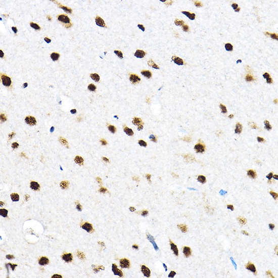
Immunohistochemistry analysis of paraffin-embedded Rat brain using SF3A3 Rabbit pAb (CAB4465) at dilution of 1:100 (40x lens). High pressure antigen retrieval performed with 0.01M Citrate buffer (pH 6.0) prior to IHC staining.
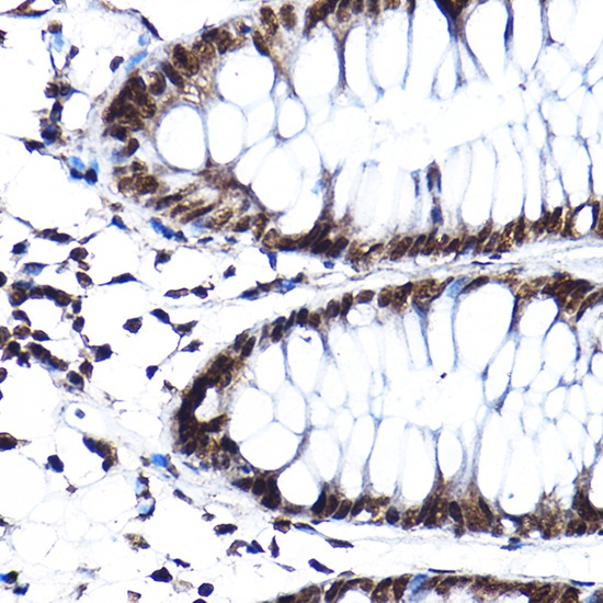
Immunohistochemistry analysis of paraffin-embedded Human colon using SF3A3 Rabbit pAb (CAB4465) at dilution of 1:100 (40x lens). High pressure antigen retrieval performed with 0.01M Citrate buffer (pH 6.0) prior to IHC staining.
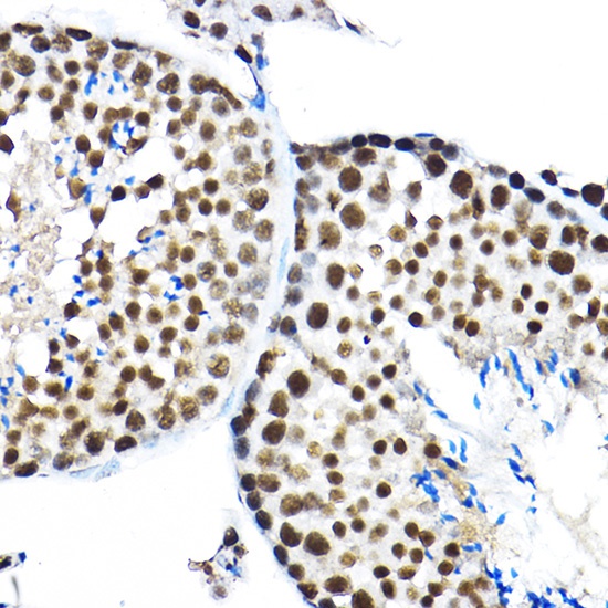
Immunohistochemistry analysis of paraffin-embedded Mouse testis using SF3A3 Rabbit pAb (CAB4465) at dilution of 1:100 (40x lens). High pressure antigen retrieval performed with 0.01M Citrate buffer (pH 6.0) prior to IHC staining.
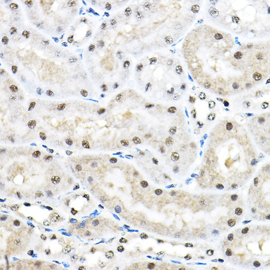
Immunohistochemistry analysis of paraffin-embedded Mouse kidney using SF3A3 Rabbit pAb (CAB4465) at dilution of 1:100 (40x lens). High pressure antigen retrieval performed with 0.01M Citrate buffer (pH 6.0) prior to IHC staining.
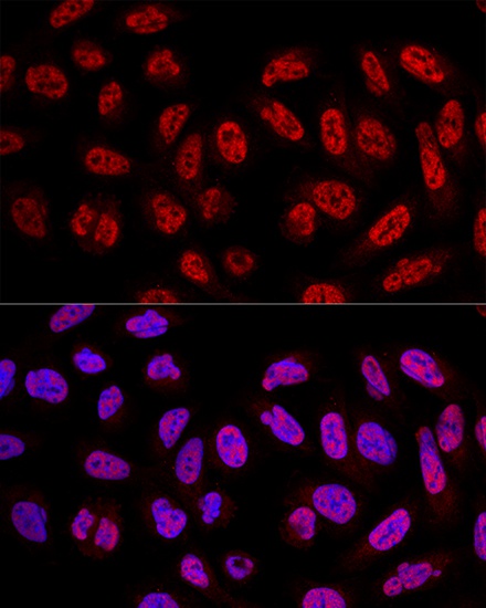
Confocal immunofluorescence analysis of U2OS cells using SF3A3 Rabbit pAb (CAB4465) at dilution of 1:100. Blue: DAPI for nuclear staining.