The SF3B3/SAP130 Monoclonal Antibody (CAB9624) is a high-quality antibody developed for reliable detection and analysis of target proteins. This antibody, generated in rabbits, exhibits high reactivity with human samples and has been validated for use in Western blot applications. By targeting the SF3B3 protein, researchers can accurately detect and analyze its expression in various cell types, making it an excellent choice for investigations in molecular biology and gene regulation.
This antibody is validated for use in WB, IHC-P, IF/ICC, ELISA applications and has demonstrated reactivity against Human, Mouse, Rat samples.
Product Name:
SF3B3/SAP130 Monoclonal Antibody
SKU:
CAB9624
Size:
20μL, 100μL
Reactivity:
Human, Mouse, Rat
Clone Number:
ARC1667
Conjugate:
Unconjugated
Immunogen:
Recombinant protein (or fragment).This information is considered to be commercially sensitive.
Recommended starting concentration is 1 μg/mL. Please optimize the concentration based on your specific assay requirements.
Synonyms:
RSE1, SAP130, SF3b130, STAF130, SF3B3/SAP130
Positive Sample:
Hep G2, Mouse testis, Rat lung
Cellular Localization:
Nucleus.
Calculated MW:
136kDa
Observed MW:
130-136kDa
This gene encodes subunit 3 of the splicing factor 3b protein complex. Splicing factor 3b, together with splicing factor 3a and a 12S RNA unit, forms the U2 small nuclear ribonucleoproteins complex (U2 snRNP). The splicing factor 3b/3a complex binds pre-mRNA upstream of the intron's branch site in a sequence independent manner and may anchor the U2 snRNP to the pre-mRNA. Splicing factor 3b is also a component of the minor U12-type spliceosome. Subunit 3 has also been identified as a component of the STAGA (SPT3-TAF(II)31-GCN5L acetylase) transcription coactivator-HAT (histone acetyltransferase) complex, and the TFTC (TATA-binding-protein-free TAF(II)-containing complex). These complexes may function in chromatin modification, transcription, splicing, and DNA repair.
Purification Method
Affinity purification
Gene ID
23450
RRID
AB_2863742
Buffer Information
Store at -20℃. Avoid freeze / thaw cycles. Buffer: PBS containing 50% glycerol and 0.05% BSA, preserved with proclin300 or sodium azide, pH 7.3.
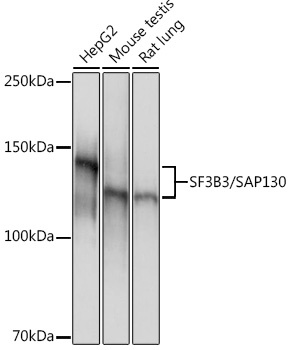
Western blot analysis of various lysates using SF3B3/SAP130 Rabbit mAb (CAB9624) at 1:1000 dilution. Secondary antibody: HRP-conjugated Goat anti-Rabbit IgG (H+L) (CABS014) at 1:10000 dilution. Lysates/proteins: 25μg per lane. Blocking buffer: 3% nonfat dry milk in TBST. Detection: ECL Basic Kit (AbGn00020). Exposure time: 1s.
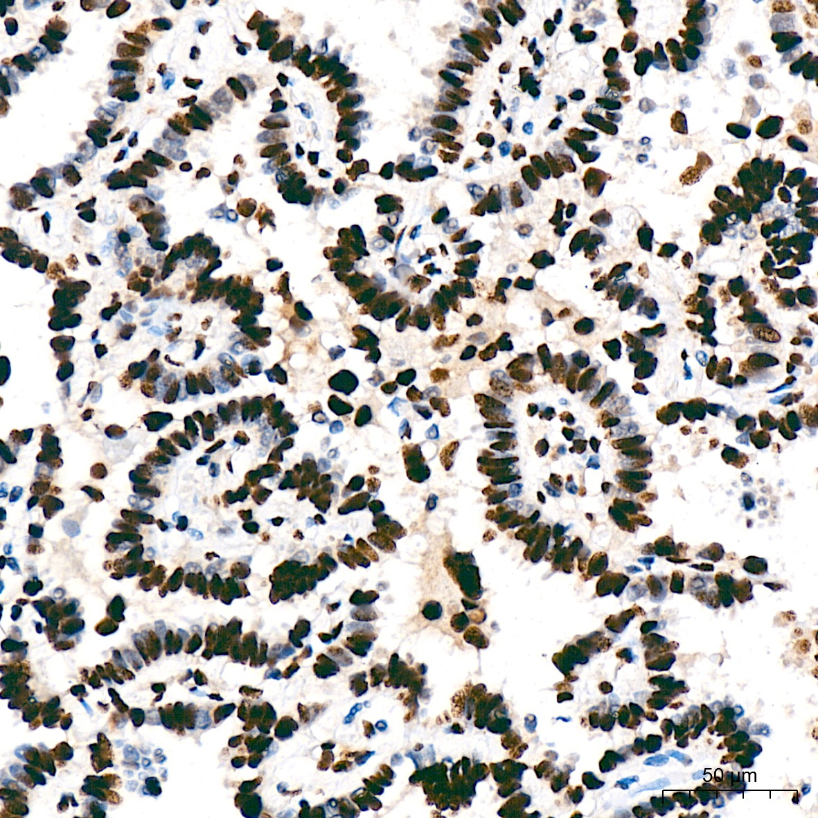
Immunohistochemistry analysis of paraffin-embedded Human lung adenocarcinoma tissue using SF3B3/SAP130 Rabbit mAb (CAB9624) at a dilution of 1:200 (40x lens). High pressure antigen retrieval was performed with 0.01 M citrate buffer (pH 6.0) prior to IHC staining.
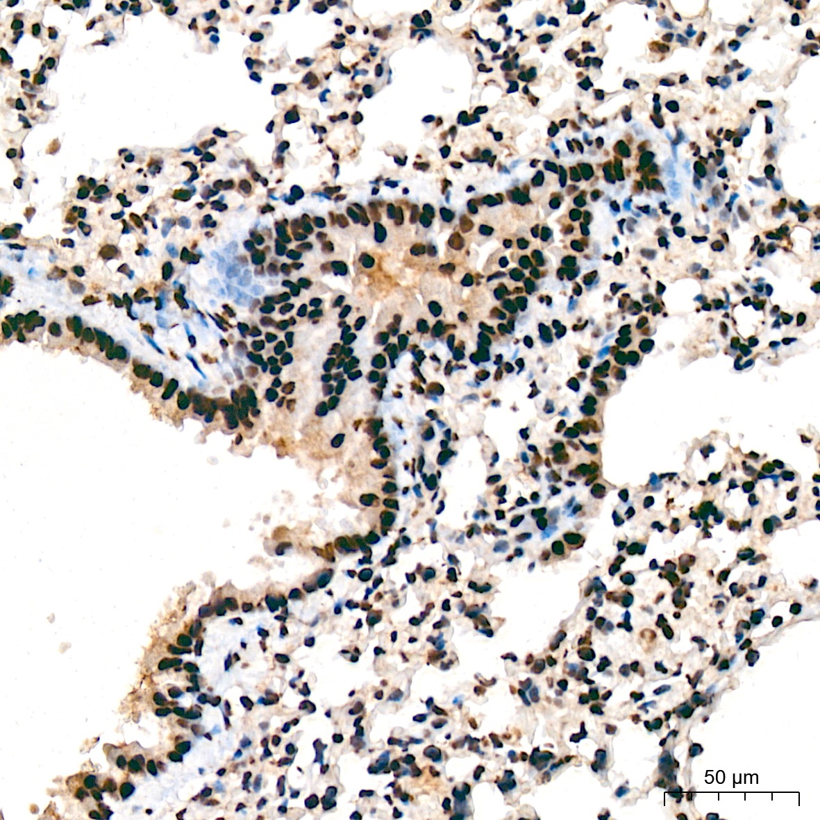
Immunohistochemistry analysis of paraffin-embedded Mouse lung tissue using SF3B3/SAP130 Rabbit mAb (CAB9624) at a dilution of 1:200 (40x lens). High pressure antigen retrieval was performed with 0.01 M citrate buffer (pH 6.0) prior to IHC staining.
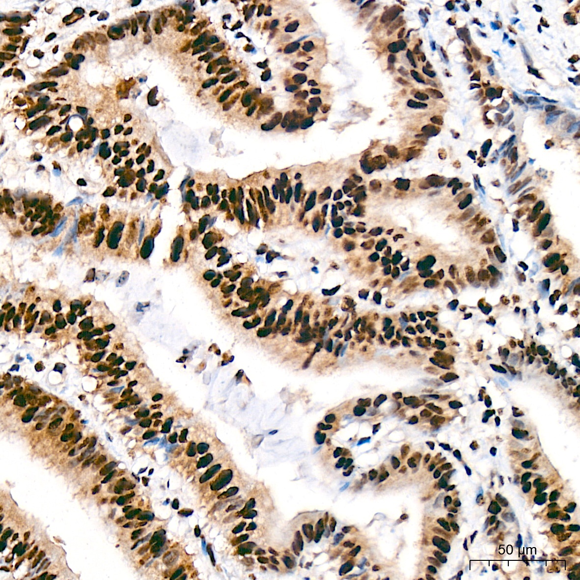
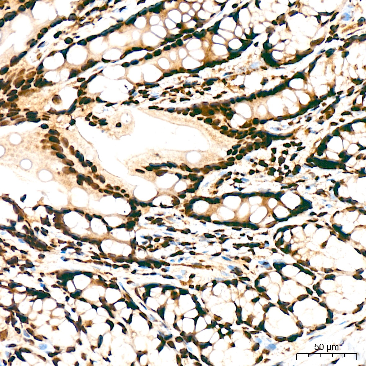
Immunohistochemistry analysis of paraffin-embedded Human colon carcinoma tissue using SF3B3/SAP130 Rabbit mAb (CAB9624) at a dilution of 1:200 (40x lens). High pressure antigen retrieval was performed with 0.01 M citrate buffer (pH 6.0) prior to IHC staining.
Immunohistochemistry analysis of paraffin-embedded Rat colon tissue using SF3B3/SAP130 Rabbit mAb (CAB9624) at a dilution of 1:200 (40x lens). High pressure antigen retrieval was performed with 0.01 M citrate buffer (pH 6.0) prior to IHC staining.
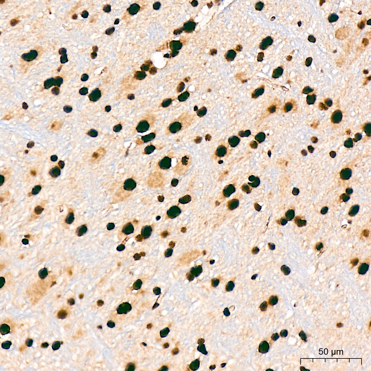
Immunohistochemistry analysis of paraffin-embedded Mouse brain tissue using SF3B3/SAP130 Rabbit mAb (CAB9624) at a dilution of 1:200 (40x lens). High pressure antigen retrieval was performed with 0.01 M citrate buffer (pH 6.0) prior to IHC staining.
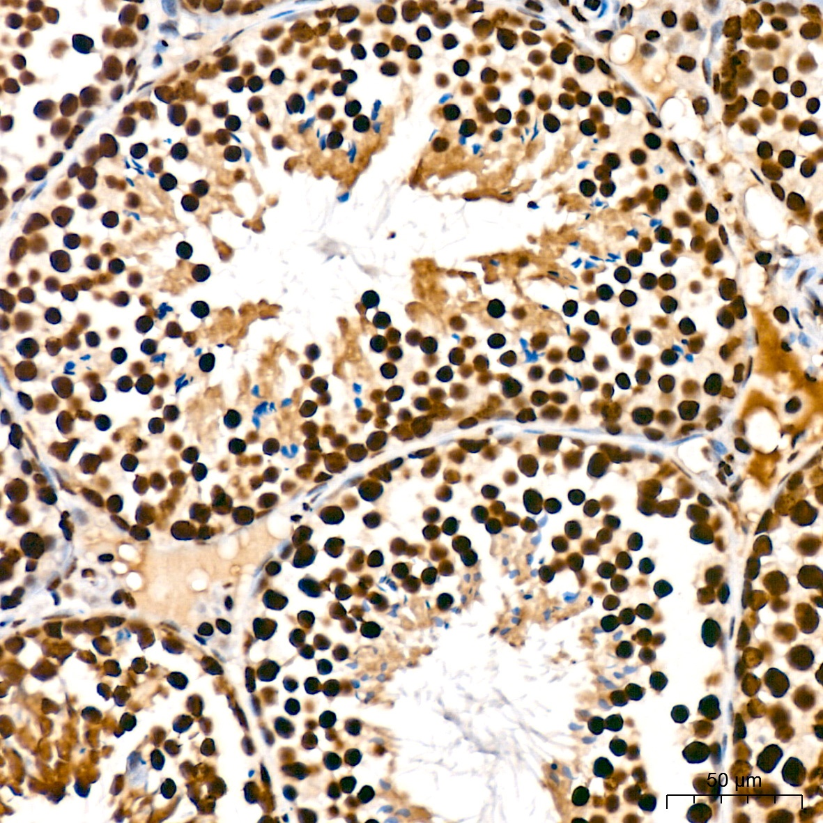
Immunohistochemistry analysis of paraffin-embedded Mouse testis tissue using SF3B3/SAP130 Rabbit mAb (CAB9624) at a dilution of 1:200 (40x lens). High pressure antigen retrieval was performed with 0.01 M citrate buffer (pH 6.0) prior to IHC staining.
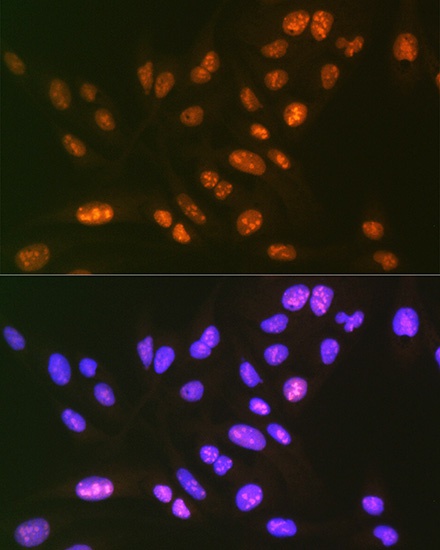
Immunofluorescence analysis of U-2 OS cells using SF3B3/SAP130 Rabbit mAb (CAB9624) at dilution of 1:100 (40x lens). Secondary antibody: Cy3-conjugated Goat anti-Rabbit IgG (H+L) (CABS007) at 1:500 dilution. Blue: DAPI for nuclear staining.


![Anti-SF3B3 [R07-2K8] Monoclonal Antibody (AGMB00714)](https://cdn11.bigcommerce.com/s-h68l9z2lnx/images/stencil/590x590/products/272003/694758/anti-sf3b3-r07-2k8-monoclonal-antibody-agmb00714__43249.1774513512.jpg?c=2 "Anti-SF3B3 [R07-2K8] Monoclonal Antibody (AGMB00714)")
![Anti-SF3B3 [R07-3F9] Monoclonal Antibody (AGMB00715)](https://cdn11.bigcommerce.com/s-h68l9z2lnx/images/stencil/590x590/products/272004/691249/anti-sf3b3-r07-3f9-monoclonal-antibody-agmb00715__32732.1774502371.jpg?c=2 "Anti-SF3B3 [R07-3F9] Monoclonal Antibody (AGMB00715)")
![Anti-SF3B3 [R05-3A-9] Monoclonal Antibody (AGMB03739)](https://cdn11.bigcommerce.com/s-h68l9z2lnx/images/stencil/590x590/products/275028/679252/anti-sf3b3-r05-3a-9-monoclonal-antibody-agmb03739__61839.1773038311.jpg?c=2 "Anti-SF3B3 [R05-3A-9] Monoclonal Antibody (AGMB03739)")