The SFRP4 Monoclonal Antibody (CAB4189) is a high-quality antibody developed for reliable detection and analysis of target proteins. This antibody, developed using rabbit monoclonal technology, exhibits high reactivity with human samples and has been validated for use in Western blot applications.SFRP4 is known for its role in modulating cell proliferation, differentiation, and migration, making it a key player in various biological processes including development, tissue homeostasis, and disease progression.
This antibody is validated for use in WB, IHC-P, IF/ICC, ELISA applications and has demonstrated reactivity against Human, Mouse, Rat samples.
Product Name:
SFRP4 Monoclonal Antibody
SKU:
CAB4189
Size:
20μL, 100μL
Reactivity:
Human, Mouse, Rat
Clone Number:
ARC0923
Conjugate:
Unconjugated
Immunogen:
Recombinant protein (or fragment).This information is considered to be commercially sensitive.
Recommended starting concentration is 1 μg/mL. Please optimize the concentration based on your specific assay requirements.
Synonyms:
PYL, FRP-4, FRPHE, FRZB-2, sFRP-4, SFRP4
Positive Sample:
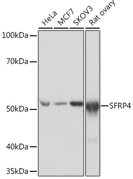
HeLa, MCF7, SKOV3, Rat ovary
Cellular Localization:
Secreted.
Calculated MW:
40kDa
Observed MW:
50kDa
Secreted frizzled-related protein 4 (SFRP4) is a member of the SFRP family that contains a cysteine-rich domain homologous to the putative Wnt-binding site of Frizzled proteins. SFRPs act as soluble modulators of Wnt signaling. The expression of SFRP4 in ventricular myocardium correlates with apoptosis related gene expression.
Purification Method
Affinity purification
Gene ID
6424
RRID
AB_2863206
Buffer Information
Store at -20℃. Avoid freeze / thaw cycles. Buffer: PBS containing 50% glycerol and 0.05% BSA, preserved with proclin300 or sodium azide, pH 7.3.
Western blot analysis of various lysates using SFRP4 Rabbit mAb (CAB4189) at 1:1000 dilution incubated overnight at 4℃. Secondary antibody: HRP-conjugated Goat anti-Rabbit IgG (H+L) (CABS014) at 1:10000 dilution. Lysates/proteins: 25μg per lane. Blocking buffer: 3% nonfat dry milk in TBST. Detection: ECL Basic Kit (AbGn00020). Exposure time: 1s.
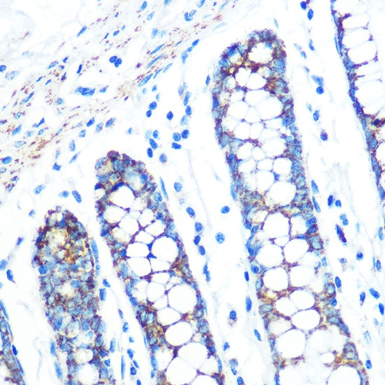
Immunohistochemistry analysis of paraffin-embedded Human colon using SFRP4 Rabbit mAb (CAB4189) at dilution of 1:100 (40x lens). Microwave antigen retrieval performed with 0.01M PBS Buffer (pH 7.2) prior to IHC staining.
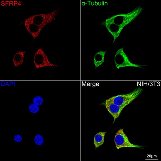
Confocal imaging of NIH/3T3 cells using SFRP4 Rabbit mAb (CAB4189,dilution 1:200) followed by a further incubation with Cy3 Goat Anti-Rabbit IgG (H+L) (CABS007,dilution 1:500)(Red).The cells were counterstained with α-Tubulin Mouse mAb (AC012, dilution 1:400) followed by incubation with ABflo® 488-conjugated Goat Anti-Mouse IgG (H+L) Ab (CABS076, dilution 1:500) (Green).DAPI was used for nuclear staining (Blue). Objective: 100x.