The SGPL1 Antibody (CAB15745) is a high-quality antibody developed for reliable detection and analysis of target proteins. This polyclonal antibody, produced in rabbits, exhibits high specificity and sensitivity towards human samples, and has been validated for use in Western blot applications.SGPL1 plays a key role in the regulation of sphingolipid levels and the generation of bioactive lipid signaling molecules, making it a crucial target for research into cancer, cardiovascular disease, and immune disorders. By binding specifically to the SGPL1 protein, this antibody enables researchers to detect and analyze SGPL1 expression in various cell types, providing valuable insights into its function and potential therapeutic applications.
This antibody is validated for use in WB, IF/ICC, ELISA applications and has demonstrated reactivity against Human, Mouse, Rat samples.
Product Name:
SGPL1 Antibody
SKU:
CAB15745
Size:
20μL, 100μL
Reactivity:
Human, Mouse, Rat
Conjugate:
Unconjugated
Immunogen:
Recombinant protein (or fragment).This information is considered to be commercially sensitive.
Recommended starting concentration is 1 μg/mL. Please optimize the concentration based on your specific assay requirements.
Synonyms:
SPL, S1PL, NPHS14, SGPL1
Positive Sample:
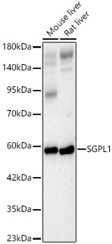
Mouse liver, Rat liver
Cellular Localization:
Endoplasmic Reticulum Membrane, Single-Pass Type Iii Membrane Protein.
Calculated MW:
64kDa
Observed MW:
60kDa
Enables sphinganine-1-phosphate aldolase activity. Involved in apoptotic signaling pathway; fatty acid metabolic process; and sphingolipid metabolic process. Located in endoplasmic reticulum. Implicated in nephrotic syndrome type 14.
Purification Method
Affinity purification
Gene ID
8879
RRID
AB_2763161
Buffer Information
Store at -20℃. Avoid freeze / thaw cycles. Buffer: PBS with 0.09% Sodium azide,50% glycerol,pH7.3.
Western blot analysis of various lysates, using SGPL1 Rabbit pAb (CAB15745) at 1:500 dilution. Secondary antibody: HRP-conjugated Goat anti-Rabbit IgG (H+L) (CABS014) at 1:10000 dilution. Lysates/proteins: 25μg per lane. Blocking buffer: 3% nonfat dry milk in TBST. Detection: ECL Basic Kit (AbGn00020). Exposure time: 60s.
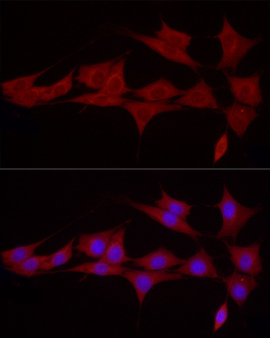
Immunofluorescence analysis of NIH/3T3 cells using SGPL1 Rabbit pAb (CAB15745) at dilution of 1:50 (40x lens). Secondary antibody: Cy3-conjugated Goat anti-Rabbit IgG (H+L) (CABS007) at 1:500 dilution. Blue: DAPI for nuclear staining.