The SH3BGRL Antibody (CAB17530) is a high-quality antibody developed for reliable detection and analysis of target proteins. This antibody, raised in rabbits, exhibits high specificity and sensitivity in detecting SH3BGR in human samples, making it ideal for use in Western blotting applications.SH3BGR, also known as a regulator of small GTPases, is involved in various cellular processes, including cell proliferation, migration, and differentiation. Dysregulation of SH3BGR has been linked to various diseases, making it a promising target for research in cancer biology, developmental biology, and cell signaling pathways.
This antibody is validated for use in WB, IF/ICC, ELISA applications and has demonstrated reactivity against Human, Mouse, Rat samples.
Product Name:
SH3BGRL Antibody
SKU:
CAB17530
Size:
20μL, 100μL
Reactivity:
Human, Mouse, Rat
Conjugate:
Unconjugated
Immunogen:
Recombinant protein (or fragment).This information is considered to be commercially sensitive.
Predicted to enable SH3 domain binding activity. Located in extracellular exosome.
Purification Method
Affinity purification
Gene ID
6451
RRID
AB_2772208
Buffer Information
Store at -20℃. Avoid freeze / thaw cycles. Buffer: PBS with 0.01% thimerosal,50% glycerol,pH7.3.
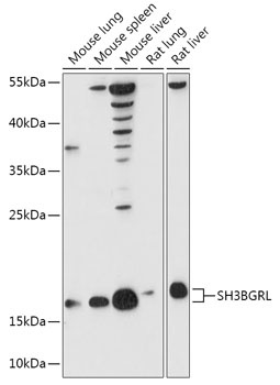
Western blot analysis of various lysates using SH3BGRL Rabbit pAb (CAB17530) at 1:1000 dilution. Secondary antibody: HRP-conjugated Goat anti-Rabbit IgG (H+L) (CABS014) at 1:10000 dilution. Lysates/proteins: 25μg per lane. Blocking buffer: 3% nonfat dry milk in TBST. Detection: ECL Basic Kit (AbGn00020). Exposure time: 90s.
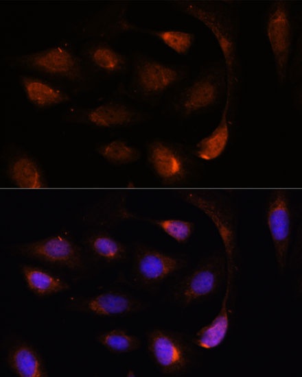
Immunofluorescence analysis of U-2 OS cells using SH3BGRL Rabbit pAb (CAB17530) at dilution of 1:100. Secondary antibody: Cy3-conjugated Goat anti-Rabbit IgG (H+L) (CABS007) at 1:500 dilution. Blue: DAPI for nuclear staining.