The SHB Monoclonal Antibody (CAB24000) is a high-quality antibody developed for reliable detection and analysis of target proteins. This monoclonal antibody, developed using hybridoma technology, provides high specificity and sensitivity for detecting SHB protein in various experimental settings.SHB is a key player in several cellular processes, including cell growth, differentiation, and survival. Its role in cancer progression and angiogenesis makes it a promising target for cancer research and drug development. By utilizing the SHB Monoclonal Antibody, researchers can investigate the function and regulation of SHB in different cell types and disease models, paving the way for new insights into therapeutic strategies for cancer and other diseases.
This antibody is validated for use in WB, ELISA applications and has demonstrated reactivity against Human, Mouse, Rat samples.
Product Name:
SHB Monoclonal Antibody
SKU:
CAB24000
Size:
20μL, 100μL
Reactivity:
Human, Mouse, Rat
Clone Number:
ARC3191
Conjugate:
Unconjugated
Immunogen:
Recombinant protein (or fragment).This information is considered to be commercially sensitive.
Sequence:
Email for sequence
Tested Applications:
WBELISA
Recommended Dilution:
WB
1:500 - 1:1000
ELISA
Recommended starting concentration is 1 μg/mL. Please optimize the concentration based on your specific assay requirements.
Enables phosphotyrosine residue binding activity. Predicted to be involved in several processes, including angiogenesis; apoptotic process; and signal transduction. Predicted to act upstream of or within several processes, including hematopoietic stem cell proliferation; negative regulation of oocyte maturation; and positive regulation of immune response. Located in cytoplasmic ribonucleoprotein granule; cytosol; and nucleoplasm.
Purification Method
Affinity purification
Gene ID
6461
Buffer Information
Store at -20℃. Avoid freeze / thaw cycles. Buffer: PBS containing 50% glycerol and 0.05% BSA, preserved with proclin300 or sodium azide, pH 7.3.
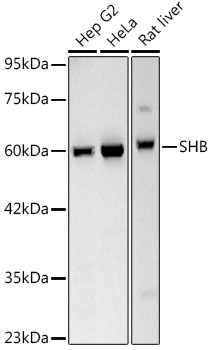
Western blot analysis of various lysates, using SHB Rabbit mAb (CAB24000) at 1:600 dilution. Secondary antibody: HRP-conjugated Goat anti-Rabbit IgG (H+L) (CABS014) at 1:10000 dilution Lysates/proteins: 25μg per lane. Blocking buffer: 3% nonfat dry milk in TBST. Detection: ECL Basic Kit (AbGn00020). Exposure time: 30s.
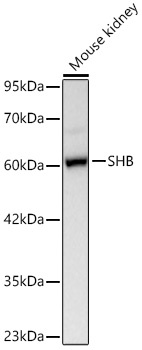
Western blot analysis of lysates from Mouse kidney using SHB Rabbit mAb (CAB24000) at 1:600 dilution incubated overnight at 4℃. Secondary antibody: HRP-conjugated Goat anti-Rabbit IgG (H+L) (CABS014) at 1:10000 dilution. Lysates/proteins: 25 μg per lane. Blocking buffer: 3% nonfat dry milk in TBST. Detection: ECL Basic Kit (AbGn00020). Exposure time: 60 s.
 at 1:600 dilution. Secondary antibody: HRP Goat Anti-Rabbit IgG (H+L) at 1:10000 dilutionLysates/proteins: 25μg per lane. Blocking buffer: 3% nonfat dry milk in TBST.")
 at 1:600 dilution. Secondary antibody: HRP Goat Anti-Rabbit IgG (H+L) at 1:10000 dilutionLysates/proteins: 25μg per lane. Blocking buffer: 3% nonfat dry milk in TBST.")


 at 1:10000 dilution. Lysates/proteins: 25ug per lane. Blocking buffer: 3% nonfat dry milk in TBST. Detection: ECL Basic Kit. Exposure time: 180s.")
 at 1:10000 dilution. Lysates/proteins: 25ug per lane. Blocking buffer: 3% nonfat dry milk in TBST. Detection: ECL Enhanced Kit. Exposure time: 45s.")