The SIVA1 Antibody (CAB6326) is a high-quality antibody developed for reliable detection and analysis of target proteins. This antibody, raised in rabbits, is highly specific and reactive with human samples, making it ideal for various research applications.SIVA1 is known to play a crucial role in regulating cell death pathways and has been implicated in cancer progression and immune system function. By targeting and binding to the SIVA1 protein, this antibody enables researchers to detect and analyze SIVA1 expression in different cell types, providing valuable insights into its role in disease pathology.
This antibody is validated for use in WB, IHC-P, ELISA applications and has demonstrated reactivity against Human, Mouse samples.
Product Name:
SIVA1 Antibody
SKU:
CAB6326
Size:
20μL, 100μL
Reactivity:
Human, Mouse
Conjugate:
Unconjugated
Immunogen:
Recombinant protein (or fragment).This information is considered to be commercially sensitive.
Recommended starting concentration is 1 μg/mL. Please optimize the concentration based on your specific assay requirements.
Synonyms:
SIVA, CD27BP, Siva-1, Siva-2, SIVA1
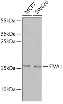
Positive Sample:
MCF7, SW620
Cellular Localization:
Cytoplasm, Nucleus.
Calculated MW:
19kDa
Observed MW:
15kDa
This gene encodes an E3 ubiquitin ligase that regulates cell cycle progression, cell proliferation and apoptosis. The N-terminus of this protein binds to the cytoplasmic tail of the CD27 antigen, a member of the tumor necrosis factor receptor (TNFR) superfamily. In response to UV radiation-induced DNA damage, this protein has been shown to mediate the ubiquitination of proliferating cell nuclear antigen (PCNA), an important step in translesion DNA synthesis.
Purification Method
Affinity purification
Gene ID
10572
RRID
AB_2766929
Buffer Information
Store at -20℃. Avoid freeze / thaw cycles. Buffer: PBS containing 50% glycerol, preserved with proclin300 or sodium azide, pH 7.3.
Western blot analysis of various lysates using SIVA1 Rabbit pAb (CAB6326) at 1:1000 dilution. Secondary antibody: HRP-conjugated Goat anti-Rabbit IgG (H+L) (CABS014) at 1:10000 dilution. Lysates/proteins: 25μg per lane. Blocking buffer: 3% nonfat dry milk in TBST. Detection: ECL Enhanced Kit (AbGn00021). Exposure time: 90s.
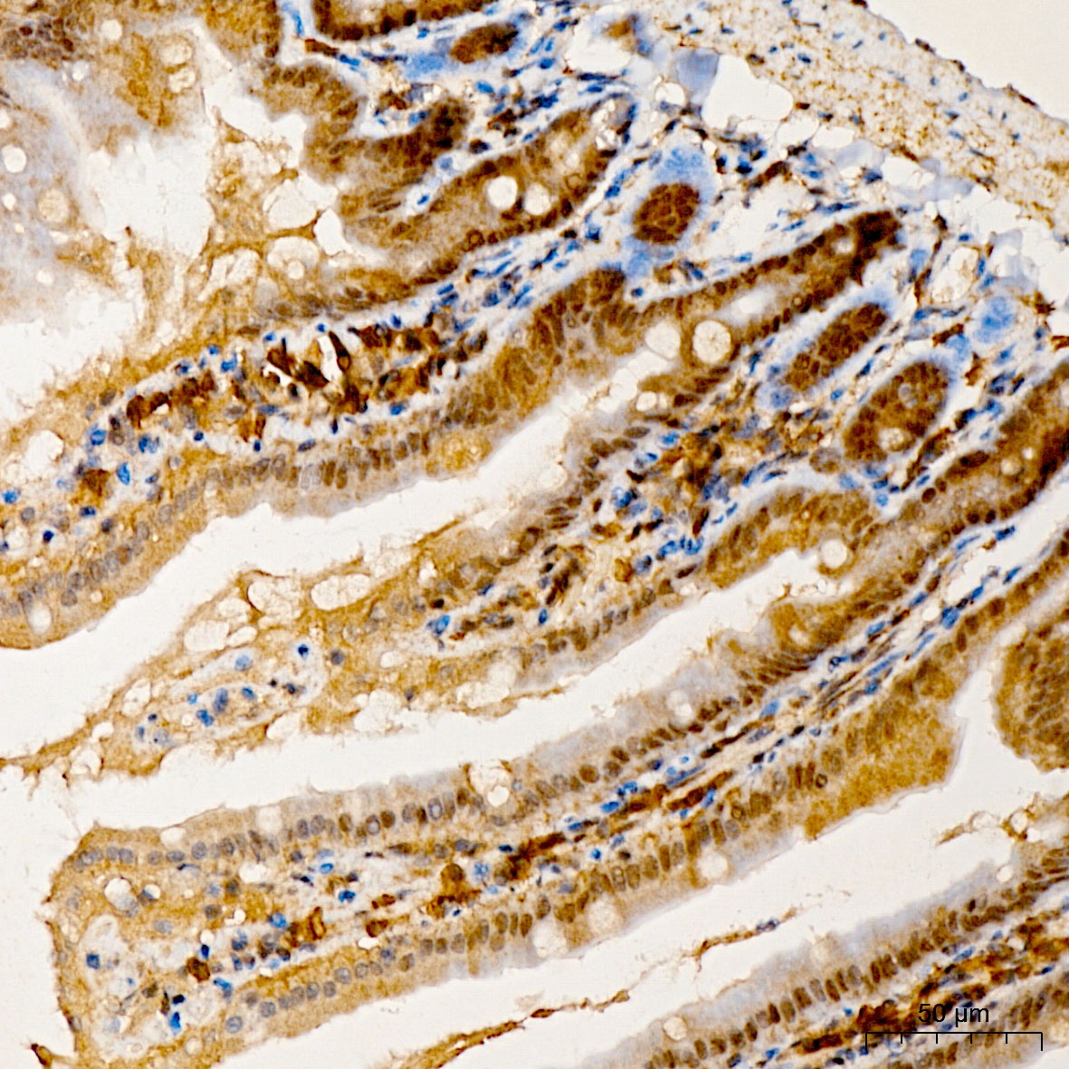
Immunohistochemistry analysis of paraffin-embedded Mouse Intestine tissue using SIVA1 Rabbit pAb (CAB6326) at a dilution of 1:200 (40x lens). High pressure antigen retrieval was performed with 0.01 M citrate buffer (pH 6.0) prior to IHC staining.