The SLAMF7 Antibody (CAB16565) is a high-quality antibody developed for reliable detection and analysis of target proteins. This antibody, produced in rabbits, exhibits high reactivity with human samples and has been validated for use in Western blot applications. By specifically binding to the SLAMF7 protein, researchers can easily detect and analyze its expression in various cell types, making it an ideal choice for studies in immunology and cancer research.SLAMF7, also known as CS1, is a key player in immune response regulation, with implications in immune cell activation and cytotoxicity.
This antibody is validated for use in WB, ELISA applications and has demonstrated reactivity against Human, Mouse samples.
Product Name:
SLAMF7 Antibody
SKU:
CAB16565
Size:
20μL, 100μL
Reactivity:
Human, Mouse
Conjugate:
Unconjugated
Immunogen:
Recombinant protein (or fragment).This information is considered to be commercially sensitive.
Recommended starting concentration is 1 μg/mL. Please optimize the concentration based on your specific assay requirements.
Synonyms:
19A, CS1, CD319, CRACC, SLAMF7
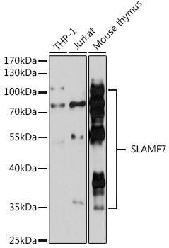
Positive Sample:
THP-1, Jurkat, Mouse thymus
Cellular Localization:
Membrane, Single-Pass Type I Membrane Protein.
Calculated MW:
37kDa
Observed MW:
35-70kDa
Enables identical protein binding activity. Predicted to be involved in adaptive immune response. Predicted to act upstream of or within regulation of natural killer cell activation. Located in endoplasmic reticulum.
Purification Method
Affinity purification
Gene ID
57823
RRID
AB_2772239
Buffer Information
Store at -20℃. Avoid freeze / thaw cycles. Buffer: PBS containing 50% glycerol, preserved with proclin300 or sodium azide, pH 7.3.
Western blot analysis of various lysates using SLAMF7 Rabbit pAb (CAB16565) at 1:3000 dilution. Secondary antibody: HRP-conjugated Goat anti-Rabbit IgG (H+L) (CABS014) at 1:10000 dilution. Lysates/proteins: 25μg per lane. Blocking buffer: 3% nonfat dry milk in TBST. Detection: ECL Basic Kit (AbGn00020). Exposure time: 90s.