The SLC1A7 Antibody (CAB10244) is a high-quality antibody developed for reliable detection and analysis of target proteins. This antibody, produced in rabbits, shows high reactivity with human samples and has been validated for use in Western blot applications. By binding specifically to the SLC1A7 protein, this antibody enables precise detection and analysis in various cell types, making it ideal for studies in the fields of biochemistry and cell biology.SLC1A7 plays a crucial role in the transport of amino acids across cell membranes, contributing to essential cellular processes such as protein synthesis and energy production.
This antibody is validated for use in WB, ELISA applications and has demonstrated reactivity against Mouse samples.
Product Name:
SLC1A7 Antibody
SKU:
CAB10244
Size:
20μL, 100μL
Reactivity:
Mouse
Conjugate:
Unconjugated
Immunogen:
Recombinant protein (or fragment).This information is considered to be commercially sensitive.
Recommended starting concentration is 1 μg/mL. Please optimize the concentration based on your specific assay requirements.
Synonyms:
AAAT, EAAT5, SLC1A7
Positive Sample:
Mouse eye
Cellular Localization:
Membrane, Multi-Pass Membrane Protein.
Calculated MW:
61kDa
Observed MW:
72kDa
Predicted to enable anion transmembrane transporter activity. Involved in neurotransmitter reuptake. Predicted to be located in plasma membrane. Predicted to be active in glutamatergic synapse. Predicted to be integral component of postsynaptic membrane and integral component of presynaptic membrane.
Purification Method
Affinity purification
Gene ID
6512
RRID
AB_2757770
Buffer Information
Store at -20℃. Avoid freeze / thaw cycles. Buffer: PBS containing 50% glycerol, preserved with proclin300 or sodium azide, pH 7.3.
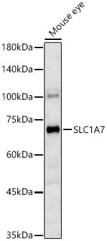
Western blot analysis of lysates from Mouse eye, using SLC1A7 Rabbit pAb (CAB10244) at 1:1000 dilution. Secondary antibody: HRP-conjugated Goat anti-Rabbit IgG (H+L) (CABS014) at 1:10000 dilution. Lysates/proteins: 25μg per lane. Blocking buffer: 3% nonfat dry milk in TBST. Detection: ECL Basic Kit (AbGn00020). Exposure time: 180s.


")


