The SLC7A1 Antibody (CAB14784) is a high-quality antibody developed for reliable detection and analysis of target proteins. This antibody, produced in rabbits, exhibits high specificity and sensitivity for human samples, making it an ideal choice for Western blot applications. By binding to the SLC7A1 protein, researchers can easily detect and analyze its expression in various cell types, facilitating investigations in fields such as metabolism, cell signaling, and cancer research.
This antibody is validated for use in WB, IHC-P, ELISA applications and has demonstrated reactivity against Human, Mouse, Rat samples.
Product Name:
SLC7A1 Antibody
SKU:
CAB14784
Size:
20μL, 100μL
Reactivity:
Human, Mouse, Rat
Conjugate:
Unconjugated
Immunogen:
Synthetic peptide. This information is considered to be commercially sensitive.
Recommended starting concentration is 1 μg/mL. Please optimize the concentration based on your specific assay requirements.
Synonyms:
ERR, ATRC1, CAT-1, HCAT1, REC1L, SLC7A1
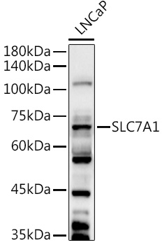
Positive Sample:
LNCaP
Cellular Localization:
Cell Membrane, Multi-Pass Membrane Protein.
Calculated MW:
68kDa
Observed MW:
68kDa
Enables L-arginine transmembrane transporter activity and L-histidine transmembrane transporter activity. Involved in amino acid transport. Located in membrane. Part of apical plasma membrane; basolateral plasma membrane; and protein-containing complex.
Purification Method
Affinity purification
Gene ID
6541
RRID
AB_2761660
Buffer Information
Store at -20℃. Avoid freeze / thaw cycles. Buffer: PBS containing 50% glycerol, preserved with proclin300 or sodium azide, pH 7.3.
Western blot analysis of lysates from LNCaP cells, using SLC7A1 Rabbit pAb (CAB14784) at 1:1000 dilution. Secondary antibody: HRP-conjugated Goat anti-Rabbit IgG (H+L) (CABS014) at 1:10000 dilution. Lysates/proteins: 25μg per lane. Blocking buffer: 3% nonfat dry milk in TBST. Detection: ECL Basic Kit (AbGn00020). Exposure time: 90s.
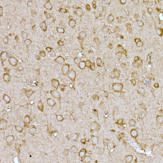
Immunohistochemistry analysis of paraffin-embedded Mouse brain using SLC7A1 Rabbit pAb (CAB14784) at dilution of 1:100 (40x lens). High pressure antigen retrieval performed with 0.01M Citrate buffer (pH 6.0) prior to IHC staining.