The Smad1/Smad5/Smad9 Polyclonal Antibody (CAB23208) is a high-quality antibody developed for reliable detection and analysis of target proteins. This antibody, produced in rabbits, is highly specific for human samples and has been validated for use in Western blot applications. By binding to SMAD1, SMAD5, and SMAD9 proteins, this antibody enables accurate detection and analysis in various cell types, making it an essential reagent for studies in developmental biology, stem cell research, and cancer biology.SMAD proteins are key components of the TGF-β signaling pathway, essential for controlling processes such as cell growth, differentiation, and apoptosis.
This antibody is validated for use in WB, IF/ICC, ELISA applications and has demonstrated reactivity against Human, Mouse samples.
Product Name:
Smad1/Smad5/Smad9 Polyclonal Antibody
SKU:
CAB23208
Size:
20μL, 100μL
Reactivity:
Human, Mouse
Conjugate:
Unconjugated
Immunogen:
Synthetic peptide. This information is considered to be commercially sensitive.
Recommended starting concentration is 1 μg/mL. Please optimize the concentration based on your specific assay requirements.
Synonyms:
Smad1/Smad5/Smad9
Positive Sample:
C2C12
Cellular Localization:
Cytoplasm, Nucleus.
Calculated MW:
52kDa
Observed MW:
52kDa
Transcriptional modulator activated by BMP (bone morphogenetic proteins) type 1 receptor kinase. SMAD1 is a receptor-regulated SMAD (R-SMAD). SMAD1/OAZ1/PSMB4 complex mediates the degradation of the CREBBP/EP300 repressor SNIP1. May act synergistically with SMAD4 and YY1 in bone morphogenetic protein (BMP)-mediated cardiac-specific gene expression.
Purification Method
Affinity purification
Gene ID
4086 4090 4093
Buffer Information
Store at -20℃. Avoid freeze / thaw cycles. Buffer: PBS containing 50% glycerol, preserved with proclin300 or sodium azide, pH 7.3.
Western blot analysis of lysates from C2C12 cells using Smad1/Smad5/Smad9 Rabbit pAb (CAB23208) at 1:1000 dilution. Secondary antibody: HRP-conjugated Goat anti-Rabbit IgG (H+L) (CABS014) at 1:10000 dilution. Lysates/proteins: 25 μg per lane. Blocking buffer: 3% nonfat dry milk in TBST. Detection: ECL Basic Kit (AbGn00020). Exposure time: 15s.
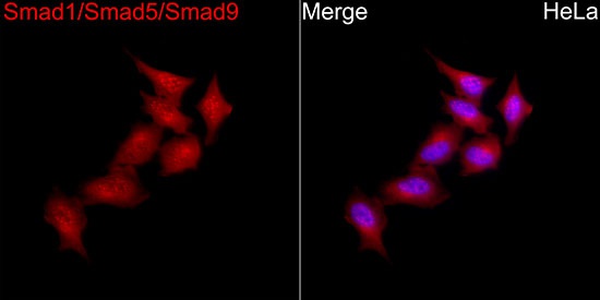
Immunofluorescence analysis of HeLa cells using Smad1/Smad5/Smad9 Rabbit pAb(CAB23208) at a dilution of 1:100 (40x lens). Secondary antibody:Cy3 Goat Anti-Rabbit IgG (H+L) (CABS007) at 1:500 dilution. Blue: DAPI for nuclear staining.
")

")


