The SMARCA1 Antibody (CAB10248) is a high-quality antibody developed for reliable detection and analysis of target proteins. This antibody, derived from rabbit serum, exhibits high specificity for human samples and has been validated for use in Western blot applications. By binding to the SMARCA1 protein, this antibody enables precise detection and analysis in a variety of cell types, making it an essential reagent for studies in molecular biology and cancer research.SMARCA1, also known as SNF2L, plays a crucial role in controlling gene expression by modulating chromatin structure.
This antibody is validated for use in WB, IHC-P, IF/ICC, IP, ELISA applications and has demonstrated reactivity against Human, Mouse, Rat samples.
Product Name:
SMARCA1 Antibody
SKU:
CAB10248
Size:
20μL, 100μL
Reactivity:
Human, Mouse, Rat
Conjugate:
Unconjugated
Immunogen:
Recombinant protein (or fragment).This information is considered to be commercially sensitive.
This gene encodes a member of the SWI/SNF family of proteins. The encoded protein is an ATPase which is expressed in diverse tissues and contributes to the chromatin remodeling complex that is involved in transcription. The protein may also play a role in DNA damage, growth inhibition and apoptosis of cancer cells. Alternative splicing results in multiple transcript variants.
Purification Method
Affinity purification
Gene ID
6594
RRID
AB_2757774
Buffer Information
Store at -20℃. Avoid freeze / thaw cycles. Buffer: PBS containing 50% glycerol, preserved with proclin300 or sodium azide, pH 7.3.
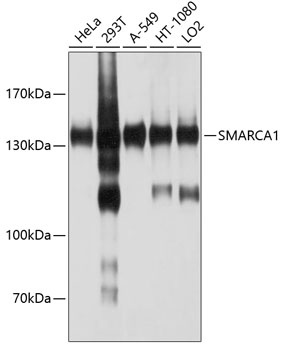
Western blot analysis of various lysates using SMARCA1 Rabbit pAb (CAB10248) at 1:1000 dilution. Secondary antibody: HRP-conjugated Goat anti-Rabbit IgG (H+L) (CABS014) at 1:10000 dilution. Lysates/proteins: 25μg per lane. Blocking buffer: 3% nonfat dry milk in TBST. Detection: ECL Basic Kit (AbGn00020). Exposure time: 15s.
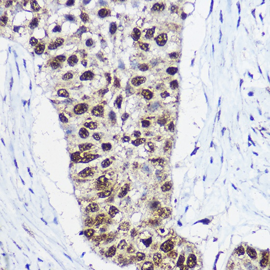
Immunohistochemistry analysis of paraffin-embedded Human lung cancer using SMARCA1 Rabbit pAb (CAB10248) at dilution of 1:100 (40x lens). High pressure antigen retrieval performed with 0.01M Citrate buffer (pH 6.0) prior to IHC staining.
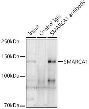
Immunoprecipitation analysis of 300 μg extracts of A-431 cells using 3 μg SMARCA1 Rabbit pAb (CAB10248). Western blot was performed from the immunoprecipitate using SMARCA1 Rabbit pAb (CAB10248) at a dilition of 1:1000.