The SMG7 Antibody (CAB15371) is a high-quality antibody developed for reliable detection and analysis of target proteins. This antibody, produced in rabbits, exhibits high reactivity towards human samples and has been validated for Western blot applications. By binding specifically to SMG7, researchers can accurately detect and analyze the protein in various cell types, making it an essential reagent for studies in molecular biology and RNA biology.SMG7 is a key player in the nonsense-mediated mRNA decay pathway, which serves as a quality control mechanism to eliminate aberrant mRNAs and regulate gene expression.
This antibody is validated for use in WB, ELISA applications and has demonstrated reactivity against Human, Mouse, Rat samples.
Product Name:
SMG7 Antibody
SKU:
CAB15371
Size:
20μL, 100μL
Reactivity:
Human, Mouse, Rat
Conjugate:
Unconjugated
Immunogen:
Recombinant protein (or fragment).This information is considered to be commercially sensitive.
Recommended starting concentration is 1 μg/mL. Please optimize the concentration based on your specific assay requirements.
Synonyms:
EST1C, SGA56M, C1orf16, SMG7
Positive Sample:
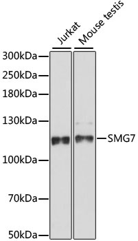
Jurkat, Mouse testis
Cellular Localization:
Cytoplasm, Nucleus.
Calculated MW:
127kDa
Observed MW:
127kDa
This gene encodes a protein that is essential for nonsense-mediated mRNA decay (NMD); a process whereby transcripts with premature termination codons are targeted for rapid degradation by a mRNA decay complex. The mRNA decay complex consists, in part, of this protein along with proteins SMG5 and UPF1. The N-terminal domain of this protein is thought to mediate its association with SMG5 or UPF1 while the C-terminal domain interacts with the mRNA decay complex. This protein may therefore couple changes in UPF1 phosphorylation state to the degradation of NMD-candidate transcripts. Alternative splicing results in multiple transcript variants encoding distinct isoforms.
Purification Method
Affinity purification
Gene ID
9887
RRID
AB_2762276
Buffer Information
Store at -20℃. Avoid freeze / thaw cycles. Buffer: PBS with 0.01% thimerosal,50% glycerol,pH7.3.
Western blot analysis of various lysates using SMG7 Rabbit pAb (CAB15371) at 1:1000 dilution. Secondary antibody: HRP-conjugated Goat anti-Rabbit IgG (H+L) (CABS014) at 1:10000 dilution. Lysates/proteins: 25μg per lane. Blocking buffer: 3% nonfat dry milk in TBST. Detection: ECL Basic Kit (AbGn00020). Exposure time: 90s.