The SOAT1 Antibody (CAB6311) is a high-quality antibody developed for reliable detection and analysis of target proteins. SOAT1, also known as acyl-coenzyme A:cholesterol acyltransferase 1, is an enzyme involved in the esterification of cholesterol in cells. This antibody, raised in rabbits, exhibits high specificity and reactivity to human samples, making it ideal for Western blot applications.SOAT1 plays a crucial role in cholesterol metabolism by converting free cholesterol into cholesterol esters, which are then stored in lipid droplets or incorporated into lipoproteins for transport.
This antibody is validated for use in WB, IF/ICC, ELISA applications and has demonstrated reactivity against Human, Mouse, Rat samples.
Product Name:
SOAT1 Antibody
SKU:
CAB6311
Size:
20μL, 100μL
Reactivity:
Human, Mouse, Rat
Conjugate:
Unconjugated
Immunogen:
Recombinant protein (or fragment).This information is considered to be commercially sensitive.
The protein encoded by this gene belongs to the acyltransferase family. It is located in the endoplasmic reticulum, and catalyzes the formation of fatty acid-cholesterol esters. This gene has been implicated in the formation of beta-amyloid and atherosclerotic plaques by controlling the equilibrium between free cholesterol and cytoplasmic cholesteryl esters. Alternatively spliced transcript variants have been found for this gene.
Purification Method
Affinity purification
Gene ID
6646
RRID
AB_2766916
Buffer Information
Store at -20℃. Avoid freeze / thaw cycles. Buffer: PBS with 0.09% Sodium azide,50% glycerol,pH7.3.
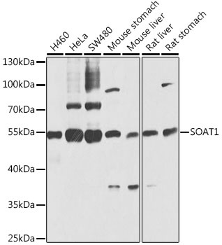
Western blot analysis of various lysates using SOAT1 Rabbit pAb (CAB6311) at 1:1000 dilution. Secondary antibody: HRP-conjugated Goat anti-Rabbit IgG (H+L) (CABS014) at 1:10000 dilution. Lysates/proteins: 25μg per lane. Blocking buffer: 3% nonfat dry milk in TBST. Detection: ECL Basic Kit (AbGn00020). Exposure time: 10s.
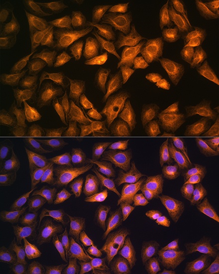
Immunofluorescence analysis of U2OS cells using SOAT1 Rabbit pAb (CAB6311) at dilution of 1:100. Secondary antibody: Cy3-conjugated Goat anti-Rabbit IgG (H+L) (CABS007) at 1:500 dilution. Blue: DAPI for nuclear staining.
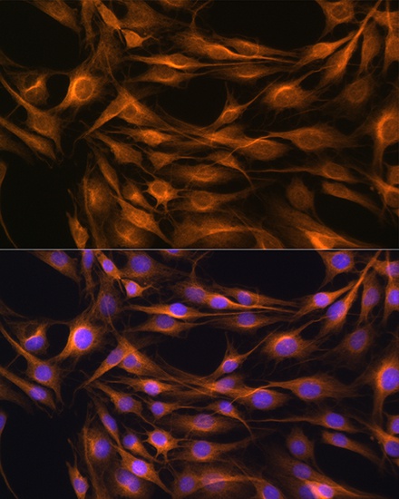
Immunofluorescence analysis of C6 cells using SOAT1 Rabbit pAb (CAB6311) at dilution of 1:100. Secondary antibody: Cy3-conjugated Goat anti-Rabbit IgG (H+L) (CABS007) at 1:500 dilution. Blue: DAPI for nuclear staining.