The SP1 Monoclonal Antibody (CAB19649) is a high-quality antibody developed for reliable detection and analysis of target proteins. This antibody, produced in rabbits, exhibits high specificity and sensitivity towards human samples, making it ideal for use in Western blot and immunohistochemistry applications.SP1 is known for its role in controlling the expression of genes involved in cell growth, differentiation, and apoptosis. Dysregulation of SP1 activity has been implicated in numerous diseases, including cancer, cardiovascular disorders, and neurological conditions.
This antibody is validated for use in WB, IHC-P, IF/ICC, ChIP, ChIP-seq, ELISA, CUT&Tag applications and has demonstrated reactivity against Human, Mouse, Rat samples.
Product Name:
SP1 Monoclonal Antibody
SKU:
CAB19649
Size:
20μL, 100μL
Reactivity:
Human, Mouse, Rat
Clone Number:
ARC0128
Conjugate:
Unconjugated
Immunogen:
Synthetic peptide. This information is considered to be commercially sensitive.
Sequence:
TTLT PIAS AASI PAGT VTVN AAQL SSMP GLQT INLS ALGT SGIQ VHPI QGLP LAIA NAPG DHGA QLGL HGAG GDGI HDDT AGGE EGEN SPDA QPQA GRRT R
Tested Applications:
WBIHC-PIF/ICCChIPChIP-seqELISACUT&Tag
Recommended Dilution:
WB
1:1000 - 1:2000
IHC-P
1:200 - 1:2000
IF/ICC
1:200 - 1:1000
ELISA
Recommended starting concentration is 1 μg/mL. Please optimize the concentration based on your specific assay requirements.
ChIP
5μg antibody for 10μg-15μg of Chromatin
ChIP-seq
1:50 - 1:100
CUT&Tag
10⁵ cells /1 μg
Synonyms:
SP1
Positive Sample:
HeLa, U-87MG, Rat liver
Cellular Localization:
Cytoplasm, Nucleus.
Calculated MW:
81kDa
Observed MW:
90kDa
The protein encoded by this gene is a zinc finger transcription factor that binds to GC-rich motifs of many promoters. The encoded protein is involved in many cellular processes, including cell differentiation, cell growth, apoptosis, immune responses, response to DNA damage, and chromatin remodeling. Post-translational modifications such as phosphorylation, acetylation, glycosylation, and proteolytic processing significantly affect the activity of this protein, which can be an activator or a repressor. Three transcript variants encoding different isoforms have been found for this gene.
Purification Method
Affinity purification
Gene ID
6667
RRID
AB_2862714
Buffer Information
Store at -20℃. Avoid freeze / thaw cycles. Buffer: PBS containing 50% glycerol and 0.05% BSA, preserved with proclin300 or sodium azide, pH 7.3.
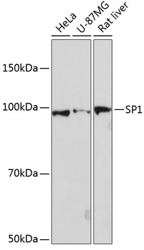
Western blot analysis of various lysates using SP1 Rabbit mAb (CAB19649) at 1:1000 dilution. Secondary antibody: HRP-conjugated Goat anti-Rabbit IgG (H+L) (CABS014) at 1:10000 dilution. Lysates/proteins: 25μg per lane. Blocking buffer: 3% nonfat dry milk in TBST. Detection: ECL Basic Kit (AbGn00020). Exposure time: 90s.
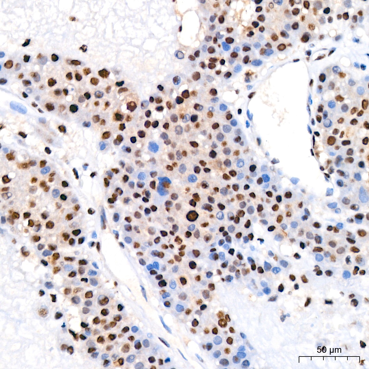
Immunohistochemistry analysis of paraffin-embedded Human liver cancer tissue using SP1 Rabbit mAb (CAB19649) at a dilution of 1:200 (40x lens). High pressure antigen retrieval performed with 0.01M Citrate buffer (pH 6.0) prior to IHC staining.
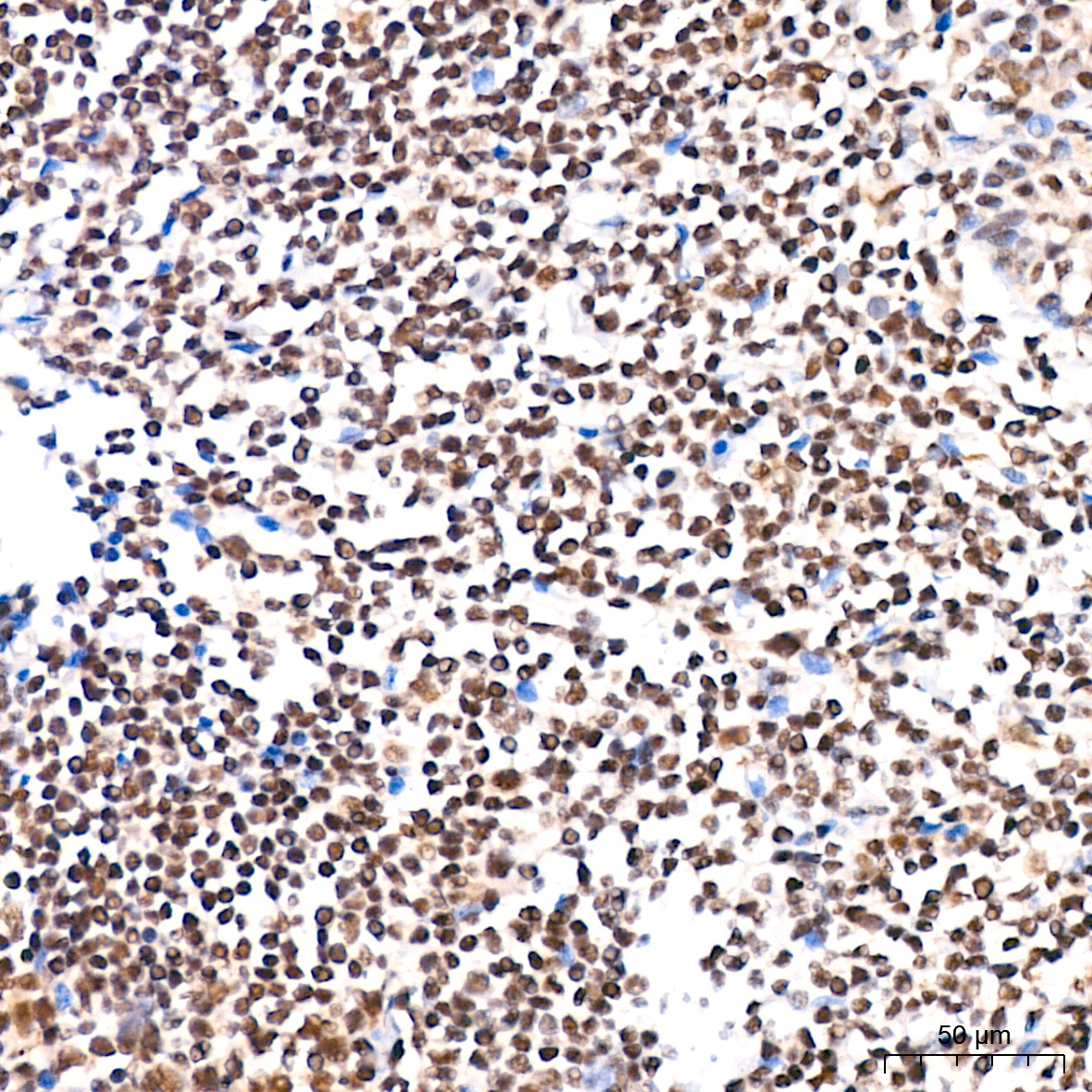
Immunohistochemistry analysis of paraffin-embedded Human tonsil tissue using SP1 Rabbit mAb (CAB19649) at a dilution of 1:200 (40x lens). High pressure antigen retrieval performed with 0.01M Citrate buffer (pH 6.0) prior to IHC staining.
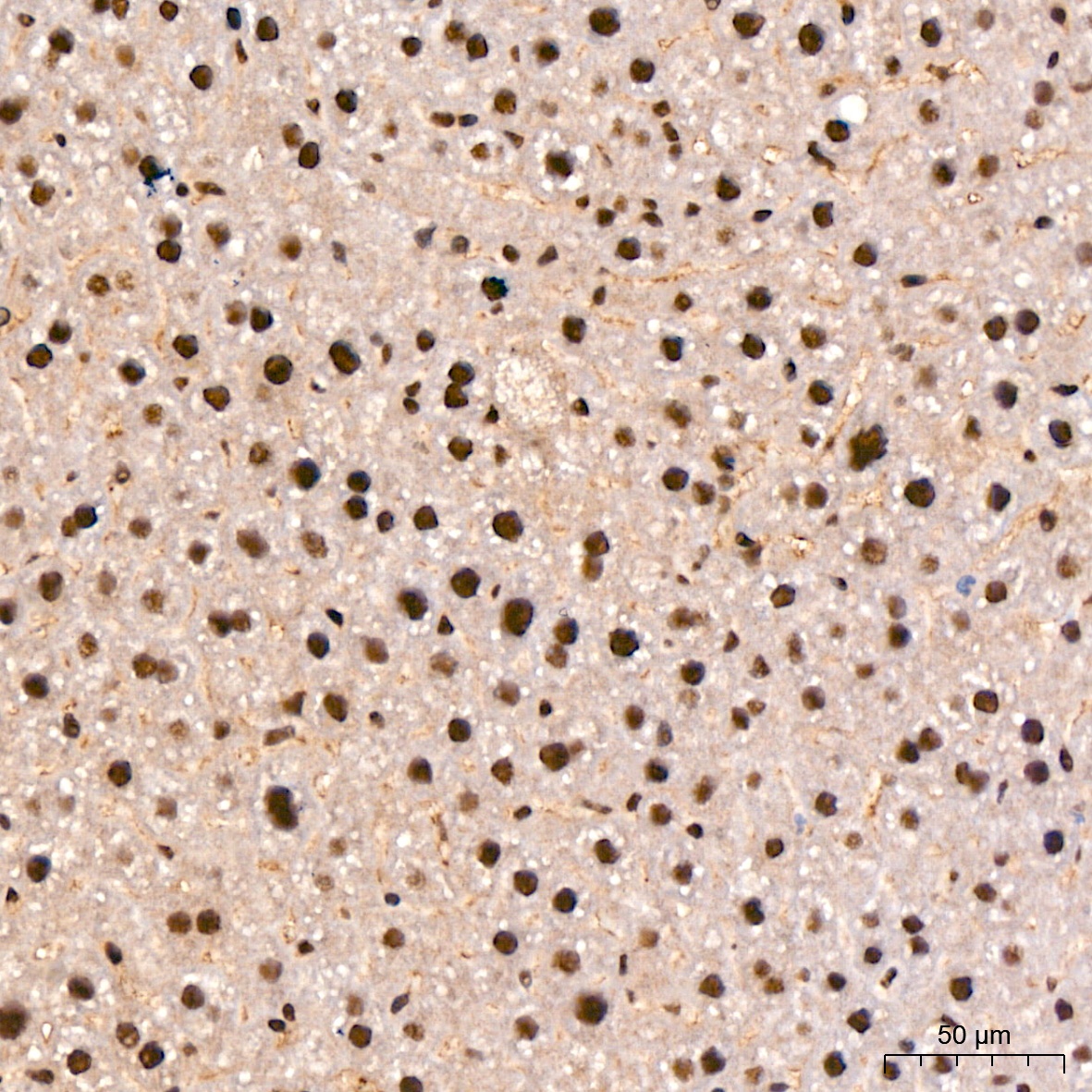
Immunohistochemistry analysis of paraffin-embedded Mouse liver tissue using SP1 Rabbit mAb (CAB19649) at a dilution of 1:200 (40x lens). High pressure antigen retrieval performed with 0.01M Citrate buffer (pH 6.0) prior to IHC staining.
Immunohistochemistry analysis of paraffin-embedded Rat spleen tissue using SP1 Rabbit mAb (CAB19649) at a dilution of 1:200 (40x lens). High pressure antigen retrieval performed with 0.01M Citrate buffer (pH 6.0) prior to IHC staining.
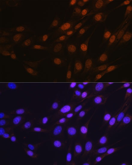
Immunofluorescence analysis of NIH-3T3 cells using SP1 Rabbit mAb (CAB19649) at dilution of 1:100 (40x lens). Secondary antibody: Cy3-conjugated Goat anti-Rabbit IgG (H+L) (CABS007) at 1:500 dilution. Blue: DAPI for nuclear staining.
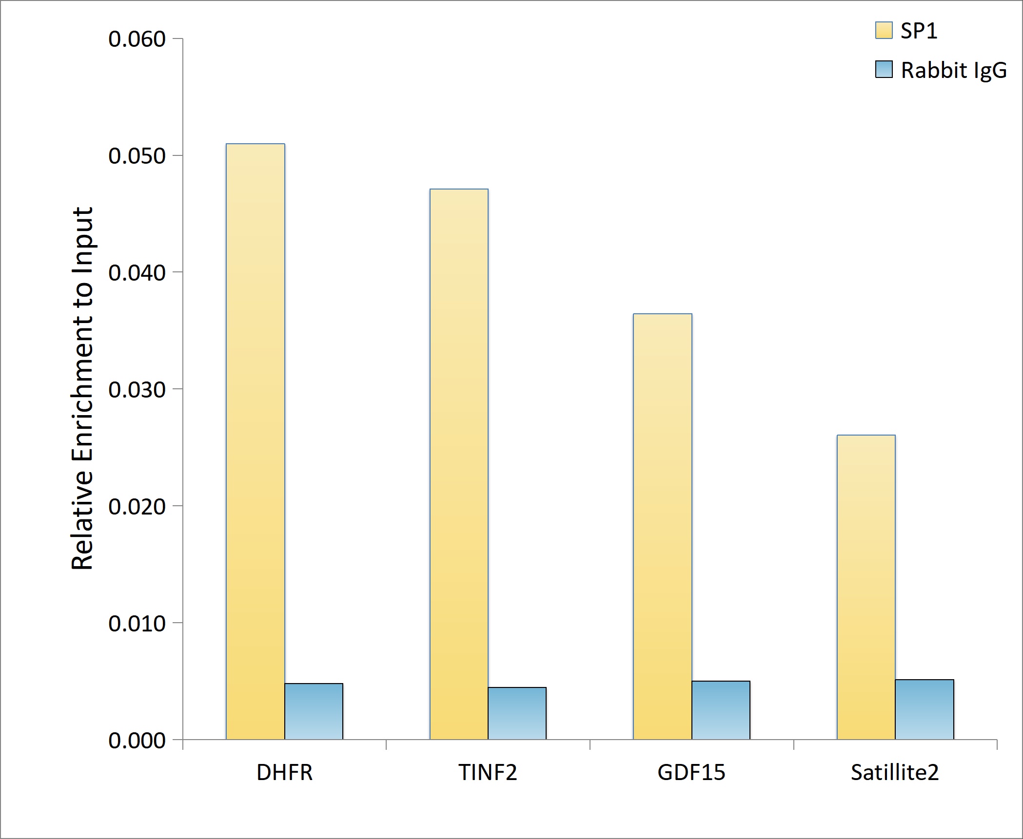
Chromatin immunoprecipitation analysis of extracts of 293T cells, using SP1 antibody (CAB19649) and rabbit IgG.The amount of immunoprecipitated DNA was checked by quantitative PCR. Histogram was constructed by the ratios of the immunoprecipitated DNA to the input.
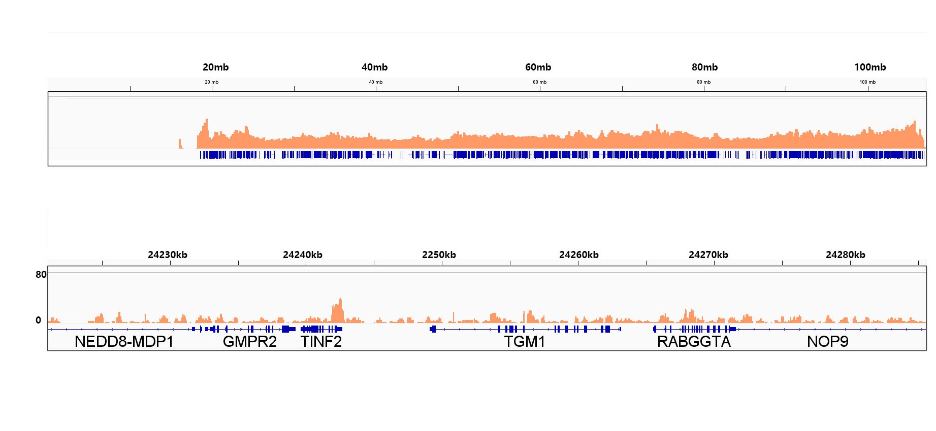
Chromatin immunoprecipitation was performed with 25 μg of cross-linked chromatin from 293T cells using 5 μg of SP1 Rabbit mAb (CAB19649). DNA libraries were prepared using Scale ssDNA-seq Lib Prep Kit for Illumina V2 (RK20228). The ChIP sequencing results indicate the enrichment pattern of SP1 across chromosome 14 (upper panel) and the genomic region encompassing TINF2, a representative gene enriched in SP1 (lower panel).
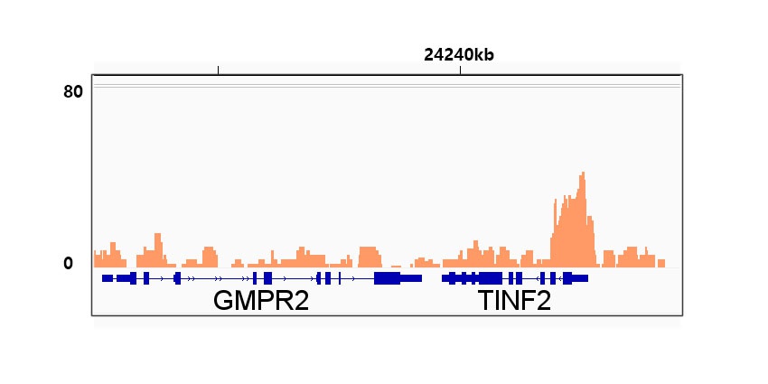
Chromatin immunoprecipitation was performed with 25 μg of cross-linked chromatin from 293T cells using 5 μg of SP1 Rabbit mAb (CAB19649). DNA libraries were prepared using Scale ssDNA-seq Lib Prep Kit for Illumina V2 (RK20228). The ChIP sequencing results indicate the enrichment pattern of SP1 in the representative genomic region surrounding TINF2 gene.